

Arginase II inhibited lipopolysaccharide-induced cell death by regulation of iNOS and Bcl-2 family proteins in macrophages
- PMID: 23639968
- PMCID: PMC3887864
- DOI: 10.1007/s10059-013-2332-7
Arginase II inhibited lipopolysaccharide-induced cell death by regulation of iNOS and Bcl-2 family proteins in macrophages
Abstract
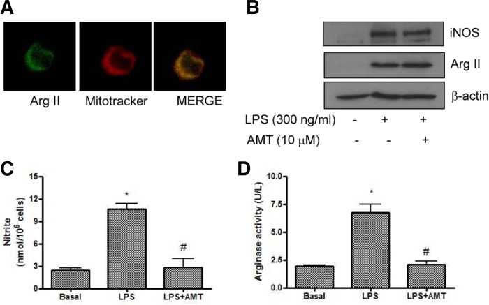
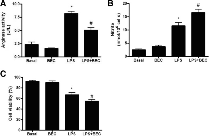
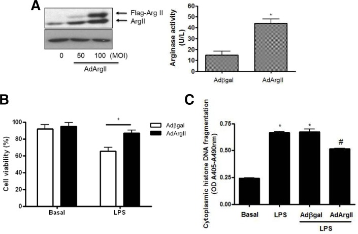
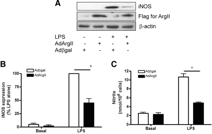
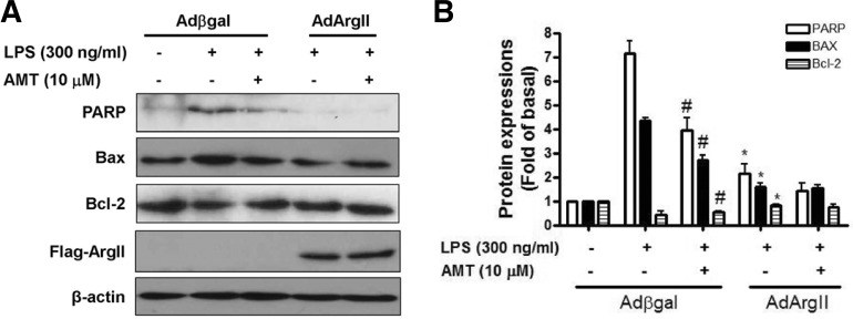
Arginase II catalyzes the conversion of arginine to urea and ornithine in many extrahepatic tissues. We investigated the protective role of arginase II on lipopolysaccharide-mediated apoptosis in the macrophage cells. Adenoviral gene transfer of full length of arginase II was performed in the murine macrophage cell line RAW264.7. The role of arginase II was investigated with cell viability, cytoplasmic histone-associated DNA fragmentation assay, arginase activity, nitric oxide production, and Western blot analysis. Arginase II is localized in mitochondria of macrophage cells, and the expression of arginase II was increased by lipopolysaccharide (LPS). LPS significantly increased cell death which was inhibited by AMT, a specific inducible nitric oxide synthase (iNOS) inhibitor. In contrast, LPS-induced cell death and nitric oxide production were increased by 2-boronoethyl-L-cysteine, a specific inhibitor of arginase. Adenoviral overexpression of arginase II significantly inhibited LPS-induced cell death and cytoplasmic histone-associated DNA fragmentation. LPS-induced iNOS expression and poly ADP-ribose polymerase cleavage were significantly suppressed by arginase II overexpression. Furthermore, arginase II overexpression resulted in a decrease in the Bax protein level and the reverse induction of Bcl-2 protein. Our data demonstrated that inhibition of NO production by arginase II may be due to arginine depletion as well as iNOS suppression though its reaction products. Moreover, arginase II plays a protective role of LPS-induced apoptosis in RAW264.7 cells.
Figures
Similar articles
-
Regulation of the urea cycle enzyme genes in nitric oxide synthesis.J Inherit Metab Dis. 1998;21 Suppl 1:59-71. doi: 10.1023/a:1005357608129. J Inherit Metab Dis. 1998. PMID: 9686345
-
MKP-1 switches arginine metabolism from nitric oxide synthase to arginase following endotoxin challenge.Am J Physiol Cell Physiol. 2007 Aug;293(2):C632-40. doi: 10.1152/ajpcell.00137.2006. Epub 2007 Apr 18. Am J Physiol Cell Physiol. 2007. PMID: 17442735
-
Arginase II downregulates nitric oxide (NO) production and prevents NO-mediated apoptosis in murine macrophage-derived RAW 264.7 cells.J Cell Biol. 1999 Feb 8;144(3):427-34. doi: 10.1083/jcb.144.3.427. J Cell Biol. 1999. PMID: 9971738 Free PMC article.
-
Regulation of nitric oxide synthesis and apoptosis by arginase and arginine recycling.J Nutr. 2007 Jun;137(6 Suppl 2):1616S-1620S. doi: 10.1093/jn/137.6.1616S. J Nutr. 2007. PMID: 17513437 Review.
-
More than just protein building blocks: how amino acids and related metabolic pathways fuel macrophage polarization.FEBS J. 2021 Jun;288(12):3694-3714. doi: 10.1111/febs.15715. Epub 2021 Feb 22. FEBS J. 2021. PMID: 33460504 Free PMC article. Review.
Cited by
-
Enhancing arginase 2 expression using target site blockers as a strategy to modulate macrophage phenotype.Mol Ther Nucleic Acids. 2022 Aug 4;29:643-655. doi: 10.1016/j.omtn.2022.08.004. eCollection 2022 Sep 13. Mol Ther Nucleic Acids. 2022. PMID: 36090747 Free PMC article.
-
Cytoplasmic localization and redox cysteine residue of APE1/Ref-1 are associated with its anti-inflammatory activity in cultured endothelial cells.Mol Cells. 2013 Nov;36(5):439-45. doi: 10.1007/s10059-013-0195-6. Epub 2013 Nov 6. Mol Cells. 2013. PMID: 24213673 Free PMC article.
-
Mitochondrial arginase-2 is essential for IL-10 metabolic reprogramming of inflammatory macrophages.Nat Commun. 2021 Mar 5;12(1):1460. doi: 10.1038/s41467-021-21617-2. Nat Commun. 2021. PMID: 33674584 Free PMC article.
-
Downregulation of L-arginine metabolism in dendritic cells induces tolerance to exogenous antigen.Int J Immunopathol Pharmacol. 2017 Mar;30(1):44-57. doi: 10.1177/0394632016678873. Epub 2017 Jan 24. Int J Immunopathol Pharmacol. 2017. PMID: 27903843 Free PMC article.
-
MicroRNA-223 alleviates lipopolysaccharide-induced PC-12 cells apoptosis and autophagy by targeting RPH1 in spinal cord injury.Int J Clin Exp Pathol. 2017 Sep 1;10(9):9223-9232. eCollection 2017. Int J Clin Exp Pathol. 2017. PMID: 31966794 Free PMC article.
References
-
- Adams JM, Cory S. The Bcl-2 protein family: arbiters of cell survival. Science. 1998;281:1322–1326. - PubMed
-
- Albina JE, Cui S, Mateo RB, Reichner JS. Nitric oxide-mediated apoptosis in murine peritoneal macrophages. J Immunol. 1993;150:5080–5085. - PubMed
-
- Boulares AH, Yakovlev AG, Ivanova V, Stoica BA, Wang G, Iyer S, Smulson M. Role of poly(ADP-ribose) polymerase (PARP) cleavage in apoptosis. Caspase 3-resistant PARP mutant increases rates of apoptosis in transfected cells. J Biol Chem. 1999;274:22932–22940. - PubMed
-
- Chao DT, Korsmeyer SJ. BCL-2 family: regulators of cell death. Annu Rev Immunol. 1998;16:395–419. - PubMed
Publication types
MeSH terms
Substances
LinkOut - more resources
Full Text Sources
Other Literature Sources
Research Materials
