. 2013 Jun 4;54(6):3880-90.
doi: 10.1167/iovs.12-10816.

Nerve growth factor promotes corneal epithelial migration by enhancing expression of matrix metalloprotease-9

Affiliations

Nerve growth factor promotes corneal epithelial migration by enhancing expression of matrix metalloprotease-9

Tomas Blanco-Mezquita et al. Invest Ophthalmol Vis Sci. .

Abstract

Purpose: Nerve growth factor (NGF) is a neuropeptide essential for the development, survival, growth, and differentiation of corneal cells. Its effects are mediated by both TrkA and p75 receptors. Clinically relevant use of NGF was introduced to treat neurotrophic ulcerations in patients. Herein, we examine the mechanisms by which NGF enhances epithelial wound healing both in vivo and in vitro.

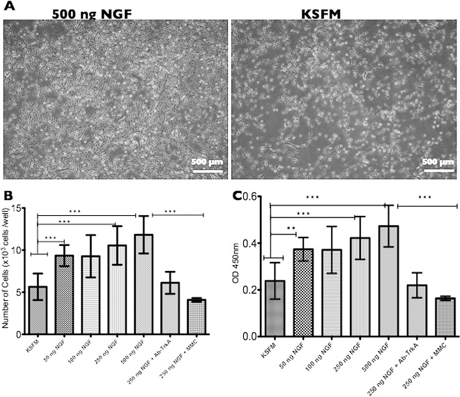
Methods: An animal model using adult hens was implemented for the in vivo experiments. Laser ablation keratectomy was performed and animals were observed for up to 7 days. Epithelial healing was measured with fluorescein. In addition, proliferation was measured using BrdU incorporation and both TrkA and matrix metalloprotease-9 (MMP-9) expression were measured by immunohistochemistry (IHC) and Western blot (WB). In vitro experiments were carried out with telomerase-immortalized human corneal epithelial cells (HCLE). The rate of proliferation was measured using a colorimetric assay and BrdU incorporation. Real-time migration was evaluated with an inverted microscope. MMP-9 expression was evaluated by immunocytochemistry (ICC), WB, zymography, and RT-PCR. Finally, beta-4 integrin (β4) expression was assessed by ICC and WB.

Results: Faster epithelial healing was observed in NGF-treated corneas compared with controls (P < 0.01). These corneas showed increased proliferation, TrkA upregulation, and enhanced MMP-9 presence (P < 0.01). In vitro, faster spreading and migration were observed in response to NGF (P < 0.01). Enhanced proliferation, as well as enhanced TrkA and MMP-9 expression, and decreased β4 levels were observed after adding NGF (P < 0.01).

Conclusions: NGF plays a major role during the epithelial healing process by promoting migration, a process that is accelerated by cell spreading. This effect is mediated by both the upregulation of MMP-9 and cleavage of β4 integrin.

Keywords: MMP-9; NGF; TrkA; corneal epithelium migration; corneal epithelium proliferation.


Figures

Figure 1
Figure 1
(A) Effect of NGF on epithelial wound closure in vivo. The time for reepithelialization was significantly reduced in the NGF group relative to both BSS and untreated controls (P < 0.01) (B). A de-organized nonconcentric but faster pattern of migration was observed in NGF corneas compared with the concentric pattern observed in both controls. (C) Corneal epithelial proliferation is shown 48 hours after surgery. More BrdU-positive cells (red) can be seen in NGF cross-sections (a) compared with both controls (b, c). Differences were statistically significant at 48 hours and 3 days (P < 0.01) (D). (E) MT stained cross-sections show a thicker epithelium with hypertrophic basal epithelial cells as well as stromal repopulation in the NGF-treated corneas (d) compared with controls that exhibit a thinner epithelium and lack of keratocytes (e, f). **P < 0.01, ***P < 0.001.
Figure 2
Figure 2
In vitro epithelial migration and wound healing. (A) The time to confluence was significantly reduced in plates supplied with 250 ng NGF relative to KSFM controls (P < 0.01). (B) The speed of migration was substantially higher (∼100 μm/h) compared with controls (40–50 μm/h) (P < 0.01). The addition of a TrkA inhibitor considerably decreased the effect of exogenous NGF. (C) Epithelial proliferation is shown by optical density 24 hours after scratching. More BrdU incorporation was observed, in a dose dependent manner, in plates supplied with NGF; however, differences were not significant compared with KSFM (P > 0.05). ***P < 0.001.
Figure 3
Figure 3
In vitro epithelial cell proliferation 24 hours after plating (3 × 10³ cells/100 μL/well). (A) Higher cell density was observed by microscope in plates supplemented with higher concentrations of NGF (500 ng) relative to KSFM controls. (B) CCK8 kit assay of cell viability 24 hours after plating is shown. The number of viable cells was significantly higher in plates supplemented with higher doses of NGF relative to KSFM (P < 0.01). A TrkA inhibitor reverted the effect of exogenous NGF and MMC compromised viability. (C) BrdU incorporation measured by ELISA. Significant differences can be observed in NGF-supplemented plates compared with KSFM controls (P < 0.01). The addition of TrkA inhibitor reverted the effects of exogenous NGF and MMC suppressed proliferation. **P < 0.01, ***P < 0.001.
Figure 4
Figure 4
Epithelial expression of the TrkA receptor. (A) Representative WB image showing a significant expression increase in TrkA in the epithelium of corneas treated with NGF, compared with both normal healthy corneas and controls. Results were normalized to β-actin and compared to baseline expression of normal healthy corneas (B). TrkA was significantly upregulated in response to exogenous NGF (P < 0.01). Increased TrkA staining can be observed in the epithelium 5 days after surgery (C). NGF-treated corneas (2) show a stronger stain compared with both controls (3, 4) and normal healthy corneas (1). Notice that NGF epithelium staining (2) is observed in all epithelial layers, whereas in controls (3, 4), staining is limited to the most basal epithelial layers. In vitro TrkA expression was measured by ICC during proliferation (D) and migration (E). Under both conditions, TrkA expression was upregulated by exogenous NGF (P < 0.01). ***P < 0.001.
Figure 5
Figure 5
MMP-9 epithelial expression. (A) MMP-9 staining increased in the epithelium 24 hours after surgery (white arrows). NGF-treated corneas (2) show stronger staining compared with controls (3, 4) and normal healthy corneas (black arrowheads; [1]). (B) Representative WB image showing the expression of active MMP-9 in the epithelium of hen corneas treated with NGF compared with both normal healthy corneas and controls. Results were normalized to β-actin and compared to the baseline expression of normal healthy corneas. MMP-9 was highly upregulated in response to exogenous NGF (P < 0.01). Only the 75-kDa band was observed. (C) Representative WB image showing the significant increase in the expression of both pro and active MMP-9 in response to NGF in culture cells after scratching. Both Pro-MMP-9 and active MMP-9 were highly increased in cells fed with NGF (P < 0.01). (D) Gelatin zymography from culture medium showing MMP-9 and MMP-2 gelatinase activity. MMP-9 gelatinase activity was increased significantly after wounding (3 and 6 hours) in aspirated medium from NGF plates with respect to controls (P < 0.01). (F) MMP-9 gene expression was measured by RT-PCR at 3, 6, and 24 hours after scratching. MMP-9 gene expression was observed more quickly and increased following exogenous NGF administration (P < 0.01). This NGF effect was diminished by a TrkA inhibitor. **P < 0.01, ***P < 0.001.
Figure 6
Figure 6
Reduction of β4 integrin in response to NGF. (A) ICC shows a drastic reduction in the β4 integrin staining in response to NGF relative to both normal and KSFM controls (P < 0.01). A TrkA inhibitor reverted the effects of exogenous NGF on β4 presence. The addition of different concentrations of a MMP-9 inhibitor reverted the effects of exogenous NGF in a dose-dependent manner. (B) A representative immunoblot showing different products as result of proteolytic cleavage of β4 integrin in response to NGF. Five different bands were detectable using a C-terminus antibody. A strong reduction in the native 205-kDa band is observed at 3 and 6 hours after scratching in the plates treated with 250 ng NGF (asterisk). A new 180-kDa band appeared at 3 and 6 hours only in cells supplemented with NGF. A third band (160 kDa) was also detectable with no significant differences between groups. However, a fourth band (100 kDa) shows up to 8-fold increase in this product 6 hours after scratching in NGF-treated cells. Finally, a fifth cleavage product (50 kDa) was increased at 6 hours in the same group. Both proteolytic activities were reverted by addiction of anti-TrkA or MMP-9 inhibitor. ***P < 0.001.

References

    1. Kawamoto K, Matsuda H. Nerve growth factor and wound healing. Prog Brain Res. 2004; 146: 369–384. - PubMed
    1. Bonini S, Lambiase A, Rama P, Caprioglio G, Aloe L. Topical treatment with nerve growth factor for neurotrophic keratitis. Ophthalmology. 2000; 107: 1347–1351; discussion 1351–1352. - PubMed
    1. Lambiase A, Manni L, Rama P, Bonini S. Clinical application of nerve growth factor on human corneal ulcer. Arch Ital Biol. 2003; 141: 141–148. - PubMed
    1. Tan MH, Bryars J, Moore J. Use of nerve growth factor to treat congenital neurotrophic corneal ulceration. Cornea. 2006; 25: 352–355. - PubMed
    1. Aloe L, Tirassa P, Lambiase A. The topical application of nerve growth factor as a pharmacological tool for human corneal and skin ulcers. Pharmacol Res. 2008; 57: 253–258. - PubMed

Publication types