Comparative Study
. 2013 Oct;58(10):2858-2867.
doi: 10.1007/s10620-013-2686-x. Epub 2013 May 3.

The Myc 3' Wnt responsive element regulates neutrophil recruitment after acute colonic injury in mice

Affiliations
Comparative Study

The Myc 3' Wnt responsive element regulates neutrophil recruitment after acute colonic injury in mice

Wesley M Konsavage Jr et al. Dig Dis Sci. 2013 Oct.

Abstract

Background: The Wnt/β-catenin pathway regulates intestinal development, homeostasis, and regeneration after injury. Wnt/β-catenin signaling drives intestinal proliferation by activating expression of the c-Myc proto-oncogene (Myc) through the Myc 3' Wnt responsive DNA element (Myc 3' WRE). In a previous study, we found that deletion of the Myc 3' WRE in mice caused increased MYC expression and increased cellular proliferation in the colon. When damaged by dextran sodium sulfate (DSS), the increased proliferative capacity of Myc 3' WRE(-/-) colonocytes resulted in a more rapid recovery compared with wild-type (WT) mice. In that study, we did not examine involvement of the immune system in colonic regeneration.

Purpose: To characterize the innate immune response in Myc 3' WRE(-/-) and WT mice during and after DSS-induced colonic injury.

Methods: Mice were fed 2.5 % DSS in their drinking water for five days to induce colonic damage and were then returned to normal water for two or four days to recover. Colonic sections were prepared and neutrophils and macrophages were analyzed by immunohistochemistry. Cytokine and chemokine levels were analyzed by probing a cytokine array with colonic lysates.

Results: In comparison with WT mice, there was enhanced leukocyte infiltration into the colonic mucosal and submucosal layers of Myc 3' WRE(-/-) mice after DSS damage. Levels of activated neutrophils were substantially increased in damaged Myc 3' WRE(-/-) colons as were levels of the neutrophil chemoattractants C5/C5a, CXCL1, and CXCL2.

Conclusion: The Myc 3' WRE regulates neutrophil infiltration into DSS-damaged colons.


Figures

Fig. 1
Fig. 1
DSS causes a greater influx of leukocytes into the colonic mucosa of Myc 3′ WRE−/− mice in comparison with WT mice. a Schematic diagram of the DSS treatment/recovery protocol. Mice were given 2.5 % DSS in their drinking water for five days and then returned to normal drinking water. Colonic tissue was collected and sectioned before treatment (pretreatment, PT), after DSS exposure (DSS), and after two (RD 2) or four (RD 4) days of returning them to normal drinking water. b H&E-stained sections of colonic tissue prepared after the indicated treatments. Each panel is a representative image from n = 3 mice examined per genotype and enlargements of the boxed regions are placed to the right of each image. Yellow arrows identify representative leukocytes in each panel. c Quantification of leukocytes in 10 fields of the colonic mucosa. Error is standard error of the mean (*p < 0.05 and **p < 0.0005)
Fig. 2
Fig. 2
DSS causes a greater influx of leukocytes into the colonic submucosa of Myc 3′ WRE−/− mice in comparison with WT mice. a H&E-stained sections of colonic tissue prepared before treatment and after five days of DSS treatment. Each panel is a representative image from n = 3 mice examined per genotype and enlargements of the boxed regions are placed to the right of each image. Yellow arrows identify representative leukocytes in each image. b Quantification of leukocytes in 10 fields of the colonic submucosa. Error is standard error of the mean (*p < 0.0005)
Fig. 3
Fig. 3
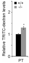
The intestinal barrier is slightly compromised in the intestines of Myc 3′ WRE−/− mice. Mice were gavaged with TRITC-dextran and fluorescence was measured in the blood 4 h later. Values detected are normalized to WT levels and n = 6 mice were examined for each genotype. Error is standard error of the mean (*p < 0.005)
Fig. 4
Fig. 4
DSS causes greater infiltration of neutrophils into the colons of Myc 3′ WRE−/− mice in comparison with WT. a Colonic sections were prepared from Myc 3′ WRE−/− and WT mice at the indicated time during the DSS protocol and neutrophils were identified by staining sections with antibodies against Ly6/G. Representative images are shown and arrows identify Ly6/G+ leukocytes. b Quantification of Ly6/G+ cells from 10 fields for each time point per mouse. c Quantification of Ly6/G+ cells in 10 fields of colonic mucosal and d submucosal layers before and after DSS treatment. In bd, n = 3 mice were examined at each time point for each genotype. Error is standard error of the mean and *p < 0.05, **p < 0.005, ***p < 0.0005
Fig. 5
Fig. 5
DSS causes greater infiltration of activated neutrophils into the colons of Myc 3′ WRE−/− mice in comparison with WT. a Relative myeloperoxidase (MPO) activity that was measured in protein homogenates from colonic tissue (n = 5 mice examined per genotype per time point; n.d. none detected). b Colonic sections were stained with Hanker–Yates reagent which detects myeloperoxidase activity. Representative images are shown and arrows indicate Hanker–Yates+ cells (n = 3 mice examined per genotype per time point). c Quantification of Hanker–Yates+ cells in 10 independent fields at the indicated times. In a, c, error is standard error of mean and *p < 0.05, **p < 0.0005
Fig. 6
Fig. 6
There is no difference in circulating neutrophils between Myc 3′ WRE−/− and WT mice. a Representative scatter plots of blood leukocytes labeled with anti-Ly6/G specific antibodies. b Average levels of circulating neutrophils reported as percentage of Ly6/G+ cells detected in purified white blood cells. Error is standard error of the mean (n = 5 mice examined per genotype). ns not significant
Fig. 7
Fig. 7
DSS causes higher expression of known neutrophil chemoattractants in the colons of Myc 3′ WRE−/− mice in comparison with WT mice. a Cytokine/chemokine arrays were probed with colonic lysates prepared from Myc 3′ WRE−/− and WT mice that were untreated or treated with DSS for five days. Signals on the blots that correspond to neutrophil chemoattractants are circled. Shown are representative blots from two independent experiments. b, c Densitometric quantification of b weakly and c highly expressed cytokines and chemokines from two independent experiments after DSS treatment. Shown is the average signal detected with error as the standard error of the mean. n.d. not detected
Fig. 8
Fig. 8
DSS induces higher levels of macrophages within colonic tissue of Myc 3′ WRE−/− mice in comparison with WT mice. a Colonic sections, collected at the indicated times, were stained with anti-F4/80 antibodies to detect macrophages. Representative images are shown and arrows depict F4/80+ cells (n = 3 mice analyzed per genotype per time point). bd Quantification of macrophages in b colonic tissue, c the colonic mucosal layer, and d the colonic submucosal layer. In each case, macrophages were tallied from 10 independent fields of view. Error is standard error of the mean and *p < 0.05, **p < 0.005

Similar articles

Cited by

References

    1. Clevers H, Nusse R. Wnt/beta-catenin signaling and disease. Cell. 2012;149:1192–1205. - PubMed
    1. Simons BD, Clevers H. Stem cell self-renewal in intestinal crypt. Exp Cell Res. 2011;317:2719–2724. - PubMed
    1. Davidson LA, Goldsby JS, Callaway ES, Shah MS, Barker N, Chapkin RS. Alteration of colonic stem cell gene signatures during the regenerative response to injury. Biochim Biophys Acta. 1822;2012:1600–1607. - PMC - PubMed
    1. Fukui T, Takeda H, Shu HJ, et al. Investigation of Musashi-1 expressing cells in the murine model of dextran sodium sulfate-induced colitis. Dig Dis Sci. 2006;51:1260–1268. - PubMed
    1. Malvin NP, Seno H, Stappenbeck TS. Colonic epithelial response to injury requires Myd88 signaling in myeloid cells. Mucosal Immunol. 2012;5:194–206. - PMC - PubMed

Publication types

MeSH terms

LinkOut - more resources