

Role of the hippocampus and orbitofrontal cortex during the disambiguation of social cues in working memory
- PMID: 23640112
- PMCID: PMC3796192
- DOI: 10.3758/s13415-013-0170-x
Role of the hippocampus and orbitofrontal cortex during the disambiguation of social cues in working memory
Abstract
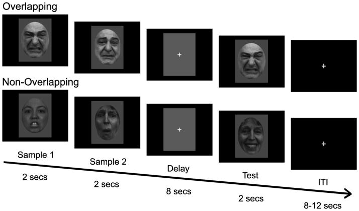
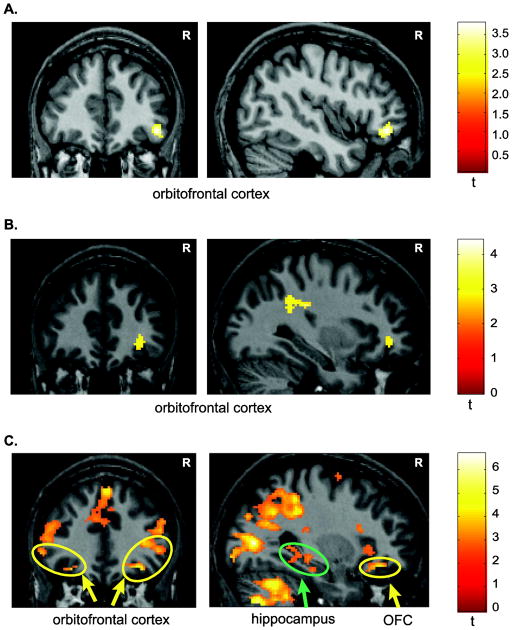
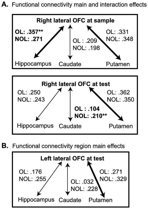
Human social interactions are complex behaviors requiring the concerted effort of multiple neural systems to track and monitor the individuals around us. Cognitively, adjusting our behavior on the basis of changing social cues such as facial expressions relies on working memory and the ability to disambiguate, or separate, the representations of overlapping stimuli resulting from viewing the same individual with different facial expressions. We conducted an fMRI experiment examining the brain regions contributing to the encoding, maintenance, and retrieval of overlapping identity information during working memory using a delayed match-to-sample task. In the overlapping condition, two faces from the same individual with different facial expressions were presented at sample. In the nonoverlapping condition, the two sample faces were from two different individuals with different expressions. fMRI activity was assessed by contrasting the overlapping and nonoverlapping conditions at sample, delay, and test. The lateral orbitofrontal cortex showed increased fMRI signal in the overlapping condition in all three phases of the delayed match-to-sample task and increased functional connectivity with the hippocampus when encoding overlapping stimuli. The hippocampus showed increased fMRI signal at test. These data suggest that lateral orbitofrontal cortex helps encode and maintain representations of overlapping stimuli in working memory, whereas the orbitofrontal cortex and hippocampus contribute to the successful retrieval of overlapping stimuli. We suggest that the lateral orbitofrontal cortex and hippocampus play a role in encoding, maintaining, and retrieving social cues, especially when multiple interactions with an individual need to be disambiguated in a rapidly changing social context in order to make appropriate social responses.
Figures
Similar articles
-
Over the river, through the woods: cognitive maps in the hippocampus and orbitofrontal cortex.Nat Rev Neurosci. 2016 Aug;17(8):513-23. doi: 10.1038/nrn.2016.56. Epub 2016 Jun 3. Nat Rev Neurosci. 2016. PMID: 27256552 Free PMC article. Review.
-
Working memory for social cues recruits orbitofrontal cortex and amygdala: a functional magnetic resonance imaging study of delayed matching to sample for emotional expressions.J Neurosci. 2008 Apr 2;28(14):3718-28. doi: 10.1523/JNEUROSCI.0464-08.2008. J Neurosci. 2008. PMID: 18385330 Free PMC article.
-
The hippocampus is functionally connected to the striatum and orbitofrontal cortex during context dependent decision making.Brain Res. 2011 Nov 14;1423:53-66. doi: 10.1016/j.brainres.2011.09.038. Epub 2011 Sep 24. Brain Res. 2011. PMID: 22000080 Free PMC article. Clinical Trial.
-
Specific and nonspecific neural activity during selective processing of visual representations in working memory.J Cogn Neurosci. 2010 Feb;22(2):292-306. doi: 10.1162/jocn.2009.21250. J Cogn Neurosci. 2010. PMID: 19400681
-
Orbitofrontal Cortex and Outcome Expectancies: Optimizing Behavior and Sensory Perception.In: Gottfried JA, editor. Neurobiology of Sensation and Reward. Boca Raton (FL): CRC Press/Taylor & Francis; 2011. Chapter 15. In: Gottfried JA, editor. Neurobiology of Sensation and Reward. Boca Raton (FL): CRC Press/Taylor & Francis; 2011. Chapter 15. PMID: 22593899 Free Books & Documents. Review.
Cited by
-
Social inference deficits in temporal lobe epilepsy and lobectomy: risk factors and neural substrates.Soc Cogn Affect Neurosci. 2015 May;10(5):636-44. doi: 10.1093/scan/nsu101. Epub 2014 Jul 25. Soc Cogn Affect Neurosci. 2015. PMID: 25062843 Free PMC article.
-
Over the river, through the woods: cognitive maps in the hippocampus and orbitofrontal cortex.Nat Rev Neurosci. 2016 Aug;17(8):513-23. doi: 10.1038/nrn.2016.56. Epub 2016 Jun 3. Nat Rev Neurosci. 2016. PMID: 27256552 Free PMC article. Review.
-
Altered resting state functional connectivity in youth with congenital heart disease operated during infancy.PLoS One. 2022 Apr 15;17(4):e0264781. doi: 10.1371/journal.pone.0264781. eCollection 2022. PLoS One. 2022. PMID: 35427374 Free PMC article.
-
Divergent effects of irradiation on brain cortical morphology in patients with nasopharyngeal carcinoma: one-year follow-up study using structural magnetic resonance imaging.Quant Imaging Med Surg. 2021 Jun;11(6):2307-2320. doi: 10.21037/qims-20-662. Quant Imaging Med Surg. 2021. PMID: 34079703 Free PMC article.
-
Inside the Developing Brain to Understand Teen Behavior From Rat Models: Metabolic, Structural, and Functional-Connectivity Alterations Among Limbic Structures Across Three Pre-adolescent Stages.Front Behav Neurosci. 2018 Sep 24;12:208. doi: 10.3389/fnbeh.2018.00208. eCollection 2018. Front Behav Neurosci. 2018. PMID: 30319367 Free PMC article.
References
-
- Barbas H, Blatt GJ. Topographically specific hippocampal projections target functionally distinct prefrontal areas in the rhesus monkey. Hippocampus. 1995;5:511–533. - PubMed
-
- Berlin HA, Rolls ET, Kischka U. Impulsivity, time perception, emotion and reinforcement sensitivity in patients with orbitofrontal cortex lesions. Brain. 2004;127(Pt 5):1108–1126. - PubMed
Publication types
MeSH terms
Substances
Grants and funding
LinkOut - more resources
Full Text Sources
Other Literature Sources
