. 2013 Jun;8(6):1028-41.
doi: 10.1038/nprot.2013.049. Epub 2013 May 2.

Derivation of extraembryonic endoderm stem (XEN) cells from mouse embryos and embryonic stem cells

Affiliations

Derivation of extraembryonic endoderm stem (XEN) cells from mouse embryos and embryonic stem cells

Kathy K Niakan et al. Nat Protoc. 2013 Jun.

Abstract

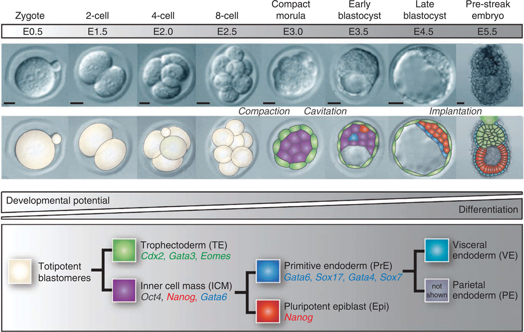
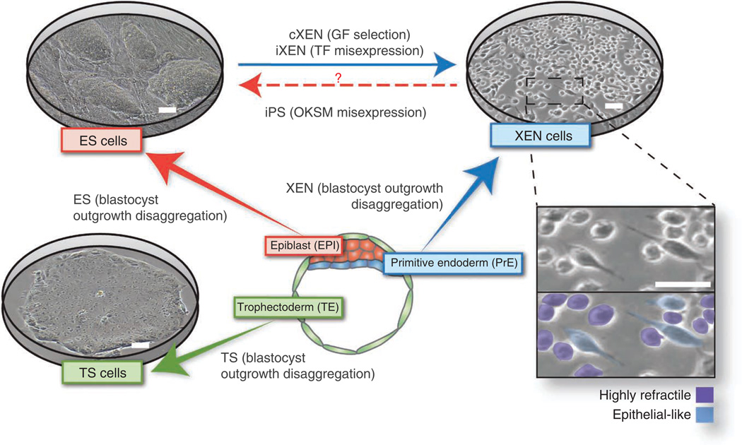
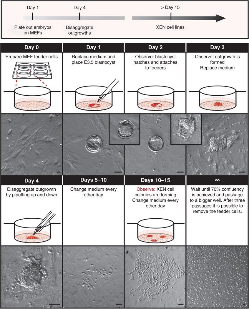
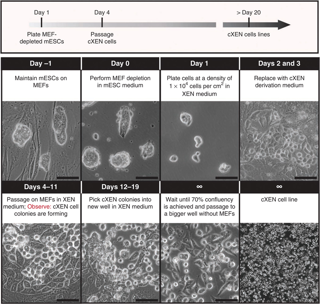
At the time of implantation in the maternal uterus, the mouse blastocyst possesses an inner cell mass comprising two lineages: epiblast (Epi) and primitive endoderm (PrE). Representative stem cells derived from these two cell lineages can be expanded and maintained indefinitely in vitro as either embryonic stem (ES) or XEN cells, respectively. Here we describe protocols that can be used to establish XEN cell lines. These include the establishment of XEN cells from blastocyst-stage embryos in either standard embryonic or trophoblast stem (TS) cell culture conditions. We also describe protocols for establishing XEN cells directly from ES cells by either retinoic acid and activin-based conversion or by overexpression of the GATA transcription factor Gata6. XEN cells are a useful model of PrE cells, with which they share gene expression, differentiation potential and lineage restriction. The robust protocols for deriving XEN cells described here can be completed within 2-3 weeks.


Figures

Figure 1
Figure 1
Overview of early embryonic development. Proper lineage segregation before implantation is ensured by two cell-fate decisions, with the first giving rise to trophectoderm and inner cell mass, and the second leading to the allocation of primitive endoderm and epiblast. Lineage-associated gene expression is noted below each cell type. After implantation, the PrE differentiates into visceral and parietal endoderm. E: embryonic day. Scale bars, 50 µm.
Figure 2
Figure 2
Stem cell types that can be derived and propagated in culture representing the three blastocyst lineages. Embryonic stem (ES) cells represent the epiblast, trophoblast stem (TS) cells represent the trophectoderm and extraembryonic endoderm (XEN) cells represent the primitive endoderm cell lineage. Heterogeneities in XEN cell morphology are indicated: highly refractile phase-bright and epithelial-like. Cognate embryo– derived stem cells retain the expression of key lineage-associated genes. GF, growth factor; iPS, induced pluripotent stem; OKSM, Oct4, Sox2, Klf4 and c-Myc. Scale bars, 100 µm.
Figure 3
Figure 3
Recovery of blastocyst-stage embryos from uteri of adult female mice. Several drops of M2 medium are prepared on the lid of a 100-mm dish. The dissected and cleaned uterine horn of a pregnant female (3.5 d.p.c.) is placed in one drop. While securing the uterine horn with forceps, the needle of a 1-ml syringe is inserted and the uterine horn is flushed with ~0.2 ml of M2 medium. The flushed blastocysts are located under high magnification and transferred to a fresh drop of M2 medium using a mouth-controlled pipette. This transfer is repeated at least twice to wash away debris, lipid drops and blood cells.
Figure 4
Figure 4
Timeline for XEN cell derivation from mouse blastocysts. Protocol for the derivation of XEN cells from blastocysts. Day 0: feeder cell–coated four-well plates are prepared. Day 1: medium is replaced and a freshly flushed E3.5 blastocyst is placed in the center of the well. Day 2: the blastocyst hatches and attaches to the feeder cells. Day 3: the blastocyst has formed an outgrowth. Medium is replaced. Day 4: the prominent outgrowth is trypsinized and disaggregated with a P20 pipette. Days 5–10: the medium is replaced every other day and the plate is observed daily. Days 10–15: XEN-like cells with stellate and refractile morphology will emerge. The medium is replaced every other day and the plate is observed daily. Day 15 and onward: After 70% confluency is attained, the XEN cells are passaged onto a well in a six-well plate and subsequently into a 100-mm dish. After three more passages they can be MEF-depleted and the XEN cell line can be frozen. Scale bars, 100 µm.
Figure 5
Figure 5
Timeline for cXEN cell derivation from mouse ES cells. Protocol for the conversion of ES cells to cXEN cells using growth factors. Day − 1: ES cells are maintained in ES medium on MEFs. Day 0: ES cells are passaged onto a pregelatinized plate for MEF depletion. Day 1: ES cells are enzymatically passaged with 0.05% (wt/vol) trypsin and plated at a density of 1 × 10⁴ cells per cm2 in standard XEN medium. Days 2 and 3: the medium is replaced with cXEN derivation medium daily (0.01–10 µM retinoic acid plus 10 ng ml− 1 activin A). Days 4–11: cells are enzymatically passaged onto MEFs in XEN medium. The medium is replaced every day or every other day depending on confluency. XEN-like cells with stellate and refractile morphology will emerge within ~5 d. Days 12–19: XEN-like cells are picked manually and placed in a MEF-coated or pregelatinized plate in XEN medium. They are passaged two more times onto pregelatinized plates and the cXEN cell line is frozen. Scale bars, 100 µm.

Similar articles

Cited by

References

    1. Schrode N, et al. Anatomy of a blastocyst: cell behaviors driving cell fate choice and morphogenesis in the early mouse embryo. Genesis. Published online; http://dx.doi.org/10.1002/dvg.22368 (25 February 2013). - DOI - PMC - PubMed
    1. Tanaka S, Kunath T, Hadjantonakis AK, Nagy A, Rossant J. Promotion of trophoblast stem cell proliferation by FGF4. Science. 1998;282:2072–2075. - PubMed
    1. Kunath T, et al. Imprinted X-inactivation in extra-embryonic endoderm cell lines from mouse blastocysts. Development. 2005;132:1649–1661. - PubMed
    1. Evans MJ, Kaufman MH. Establishment in culture of pluripotential cells from mouse embryos. Nature. 1981;292:154–156. - PubMed
    1. Martin GR. Isolation of a pluripotent cell line from early mouse embryos cultured in medium conditioned by teratocarcinoma stem cells. Proc. Natl. Acad. Sci. USA. 1981;78:7634–7638. - PMC - PubMed

Publication types