. 2013 Jun 11;108(11):2281-90.
doi: 10.1038/bjc.2013.206. Epub 2013 May 2.

Myeloid-derived suppressor cells have a central role in attenuated Listeria monocytogenes-based immunotherapy against metastatic breast cancer in young and old mice

Affiliations

Myeloid-derived suppressor cells have a central role in attenuated Listeria monocytogenes-based immunotherapy against metastatic breast cancer in young and old mice

D Chandra et al. Br J Cancer. .

Abstract

Background: Myeloid-derived suppressor cells (MDSCs) are present in large numbers in blood of mice and humans with cancer, and they strongly inhibit T-cell and natural killer (NK) cell responses, at young and old age. We found that a highly attenuated bacterium Listeria monocytogenes (Listeria(at))-infected MDSC and altered the immune-suppressing function of MDSC.

Methods: Young (3 months) and old (18 months) BALB/cByJ mice with metastatic breast cancer (4T1 model) were immunised with Listeria(at) semi-therapeutically (once before and twice after tumour development), and analysed for growth of metastases and primary tumour, in relation to MDSC-, CD8 T-cell and NK cell responses.

Results: We found that Listeria(at)-infected MDSC, which delivered Listeria(at) predominantly to the microenvironment of metastases and primary tumours, where they spread from MDSC into tumour cells (infected tumour cells will ultimately become a target for Listeria-activated immune cells). Immunotherapy with Listeria(at) significantly reduced the population of MDSC in blood and primary tumours, and converted a remaining subpopulation of MDSC into an immune-stimulating phenotype producing IL-12, in correlation with significantly improved T-cell and NK cell responses to Listeria(at) at both ages. This was accompanied with a dramatic reduction in the number of metastases and tumour growth at young and old age.

Conclusions: Although preclinical studies show that immunotherapy is less effective at old than at young age, our study demonstrates that Listeria(at)-based immunotherapy can be equally effective against metastatic breast cancer at both young and old age by targeting MDSC.


Figures
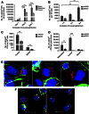
Figure 1
Figure 1
Survival and multiplication of Listeriaat in the TME, normal tissues, and MDSC.(A) Listeriaat multiplied in metastases and primary tumour but not in spleen (tumour-free tissue) in vivo. 4T1 tumour-bearing mice were injected once with 0.5 × 10⁷ Listeriaat and analysed for the number of Listeriaat in primary tumour, metastases, and spleen at different time intervals. n=3 mice per group. Mice were individually analysed and the results were averaged. The graph is a representative of three experiments. (B) Listeriaat infected both types of MDSC but multiplies in the mMDSC population in vitro. Splenic MDSC of tumour-bearing mice were isolated and cultured with Listeriaat at a 1 : 10 ratio for an hour, and then gentamicin was added, and terminated at different time points. The infected cells were lysed in water and plated onto LB agar to determine the number of Listeriaat CFU the next day. n=3 mice per group. Results of two experiments were averaged. Analysis of variance (ANOVA, one-way) *P<0.05, **P<0.01, ***P<0.001, ****P<0.0001 is significant. (C) The number of Listeriaat CFU in tumour MDSC was much higher than in splenic MDSC. Tumour-bearing mice were injected with Listeriaat as in A. The next day, mice were euthanised and MDSC were isolated from tumours and spleens, and analysed the MDSCs for their number of Listeriaat CFU. n=3 mice. Mice were individually analysed and the results were averaged, representative of two experiments. Unpaired t-test P<0.05 is statistically significant. (D) mMDSC delivered Listeriaat predominantly to the tumour microenvironment. mMDSC and gMDSC isolated from spleens of tumour-bearing mice were infected in vitro with Listeriaat, and injected (107 cells) into the tail vein of tumour-bearing mice. Next day, the number of Listeriaat CFU was determined in the primary tumours, metastases, and spleens. Mice were individually analysed and the results were averaged, representative of two experiments. n=5 mice per group ANOVA (one-way) *P<0.05, **P<0.01, ***P<0.001, ****P<0.0001 is significant. The error bars on all graphs represent the s.e.m. (E) Listeriaat spread from MDSC to tumour cells. Isolated splenic MDSC from tumour-bearing mice were infected with Listeriaat for 1 h, treated with gentamicin, and then cultured with 4T1 tumour cells (that has never been exposed to Listeriaat before) in vitro in the presence of gentamicin, and the cultures were finally terminated at various time points. Left: Listeriaat migrating from MDSC into tumour cell (after 6 h). Middle: Listeriaat in cytoplasm of dying tumour cell (after 12 h). Right: Listeriaat in cytoplasm of dying tumour cell (after 24 h). Listeriaat are red (Cy-3) and nuclei blue (DAPI), and cytoplasm green (actin staining). (F) Listeriaat-infected MDSC at young and old age. Splenic MDSC were isolated from young and old tumour-bearing mice and infected with Listeriaat as described in E. Left: Listeriaat in the cytoplasm of MDSCs from young mice. Right: Listeriaat in the cytoplasm of MDSC from old mice.
Figure 2
Figure 2
Listeriaat immunisations significantly reduced the percentage of gMDSC in blood of both young and old mice.Mice were immunised with Listeriaat on days 0, 7, and 14, and challenged with 4T1 tumour cells on day 4. All mice were euthanised on day 16 and analysed by flow cytometry for the total MDSC population (CD11b+Gr1+), the gMDSC population (CD11b+Gr1high), and the mMDSC population (CD11b+Gr1low), in blood of young and old mice. All MDSCs were gated within the total live leukocyte population of the blood. Graphs are the average of three experiments (mice were individually analysed). n=5 mice per group. Unpaired t-test. *P<0.05, **P<0.01, ***P<0.001, ****P<0.0001 is significant. The error bars represent the s.e.m. A representative example of gating MDSC of young and old mice is shown in Supplementary Figure 6 of the Supplementary Information. In addition, a representative example of absolute numbers of MDSC, gMDSC, and mMDSC in blood of young and old mice is shown in Supplementary Figure 7 of the Supplementary Information.
Figure 3
Figure 3
Listeriaat immunisations significantly reduced the percentage of gMDSC in primary tumours of both young and old mice.All mice were immunised with Listeriaat, challenged with 4T1 tumour cells, and euthanised as described in Figure 2. The total MDSC population (CD11b+Gr1+), as well as the gMDSC population (CD11b+Gr1high) and the mMDSC population (CD11b+Gr1low), were gated within the total live CD45+ population of the tumour cell suspension, and analysed by flow cytometry. Graphs are the average of three experiments (mice were individually analysed). n=5 mice per group. Unpaired t-test. *P<0.05, **P<0.01, ***P<0.001, ****P<0.0001 is significant. The error bars represent the s.e.m. A representative example of gating MDSC of young and old mice is shown in Supplementary Figure 6 of the Supplementary Information.
Figure 4
Figure 4
Listeriaat immunisations significantly increased the production of IL-12 by MDSC in blood of both young and old mice.All mice were immunised with Listeriaat, challenged with 4T1 tumour cells, and euthanised as described in Figure 2. The gMDSC and mMDSC were gated in the blood as shown in Supplementary Figure 6 of the Supplementary Information, and analysed for the intracellular production of IL-12 by flow cytometry (A). n=3–5 mice per group. Mice were individually analysed and the results of three experiments were averaged. Unpaired t-test. *P<0.05, **P<0.01, ***P<0.001, ****P<0.0001 is significant. The error bars represent the s.e.m. The IL-12 production by gMDSC and mMDSC was also analysed in vitro. For this purpose, gMDSC and mMDSC were isolated from spleens of tumour-bearing mice as described in Figure 1, infected with Listeriaat at various ratio's (gMDSC or mMDSC/Listeriaat=1:0, 0.01, and 0.1) for 1 h, then cultured for 72 h in the presence of gentamicin, and then analysed for the production of IL-12 in the culture supernatant by ELISA (B). This experiment was performed two times and the results were averaged. Unpaired t-test P<0.5 is statistically significant. The error bars represent the s.e.m.
Figure 5
Figure 5
Listeriaat-activated CD8 T cells and NK cells at both young and old age.All mice were immunised with Listeriaat, challenged with 4T1 tumour cells, and euthanised as described in Figure 2. Splenocytes were isolated from tumour-bearing young and old mice, pooled in each group, re-stimulated with Listeriaat, and analysed for the number of IFN-γ-producing CD8 T cells and NK cells by ELISPOT. CD8 T cells and NK cells were depleted by magnetic bead technique. The number of IFN-γ-producing spots was determined per 200 000 splenocytes. n=5 mice per group. This experiment was performed two times and the results were averaged. Unpaired t-test. *P<0.05, **P<0.01, ***P<0.001, ****P<0.0001 is significant. The error bars represent the s.e.m.
Figure 6
Figure 6
Listeriaat immunisations significantly reduced the number of metastases and tumour growth in both young and old mice.All mice were immunised with Listeriaat, challenged with 4T1 tumour cells, and euthanised as described in Figure 2. The number of metastases (A) and tumour weight (B) was determined. n=5 mice per group. This experiment was performed three times and the results were averaged. Unpaired t-test. *P<0.05, **P<0.01, ***P<0.001, ****P<0.0001 is significant. The error bars represent the s.e.m.

References

    1. Aslakson CJ, Miller FR. Selective events in the metastatic process defined by analysis of the sequential dissemination of subpopulations of a mouse mammary tumor. Cancer Res. 1992;52 (6:1399–1405. - PubMed
    1. Castro F, Leal B, Denny A, Bahar R, Lampkin S, Reddick R, Lu S, Gravekamp C. Vaccination with Mage-b DNA induces CD8 T-cell responses at young but not old age in mice with metastatic breast cancer. Br J Cancer. 2009;101 (8:1329–1337. - PMC - PubMed
    1. De Backer O, Verheyden AM, Martin B, Godelaine D, De Plaen E, Brasseur R, Avner P, Boon T. Structure, chromosomal location, and expression pattern of three mouse genes homologous to the human MAGE genes. Genomics. 1995;28 (1:74–83. - PubMed
    1. Diaz-Montero CM, Salem ML, Nishimura MI, Garrett-Mayer E, Cole DJ, Montero AJ. Increased circulating myeloid-derived suppressor cells correlate with clinical cancer stage, metastatic tumor burden, and doxorubicin-cyclophosphamide chemotherapy. Cancer Immunol Immunother. 2009;58 (1:49–59. - PMC - PubMed
    1. Extermann M. Basic assessment of the older cancer patient. Curr Treat Options Oncol. 2011;12 (3:276–285. - PubMed

Publication types

MeSH terms