

TGF-β1 exposure induces epithelial to mesenchymal transition both in CSCs and non-CSCs of the A549 cell line, leading to an increase of migration ability in the CD133+ A549 cell fraction
- PMID: 23640462
- PMCID: PMC3674353
- DOI: 10.1038/cddis.2013.144
TGF-β1 exposure induces epithelial to mesenchymal transition both in CSCs and non-CSCs of the A549 cell line, leading to an increase of migration ability in the CD133+ A549 cell fraction
Expression of concern in
-
Editorial Expression of Concern to: TGF-β1 exposure induces epithelial to mesenchymal transition both in CSCs and non-CSCs of the A549 cell line, leading to an increase of migration ability in the CD133+ A549 cell fraction.Cell Death Dis. 2025 Jul 2;16(1):484. doi: 10.1038/s41419-025-07780-0. Cell Death Dis. 2025. PMID: 40603307 Free PMC article. No abstract available.
Abstract
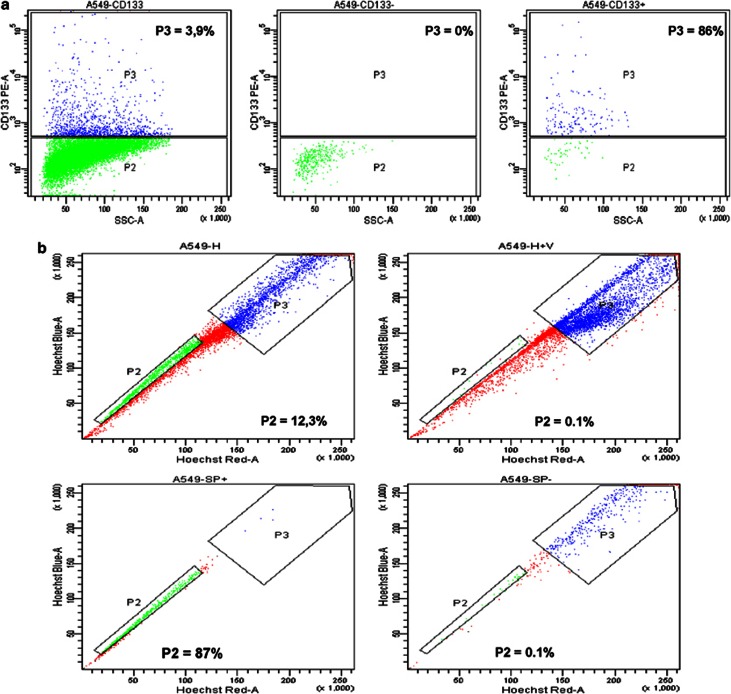
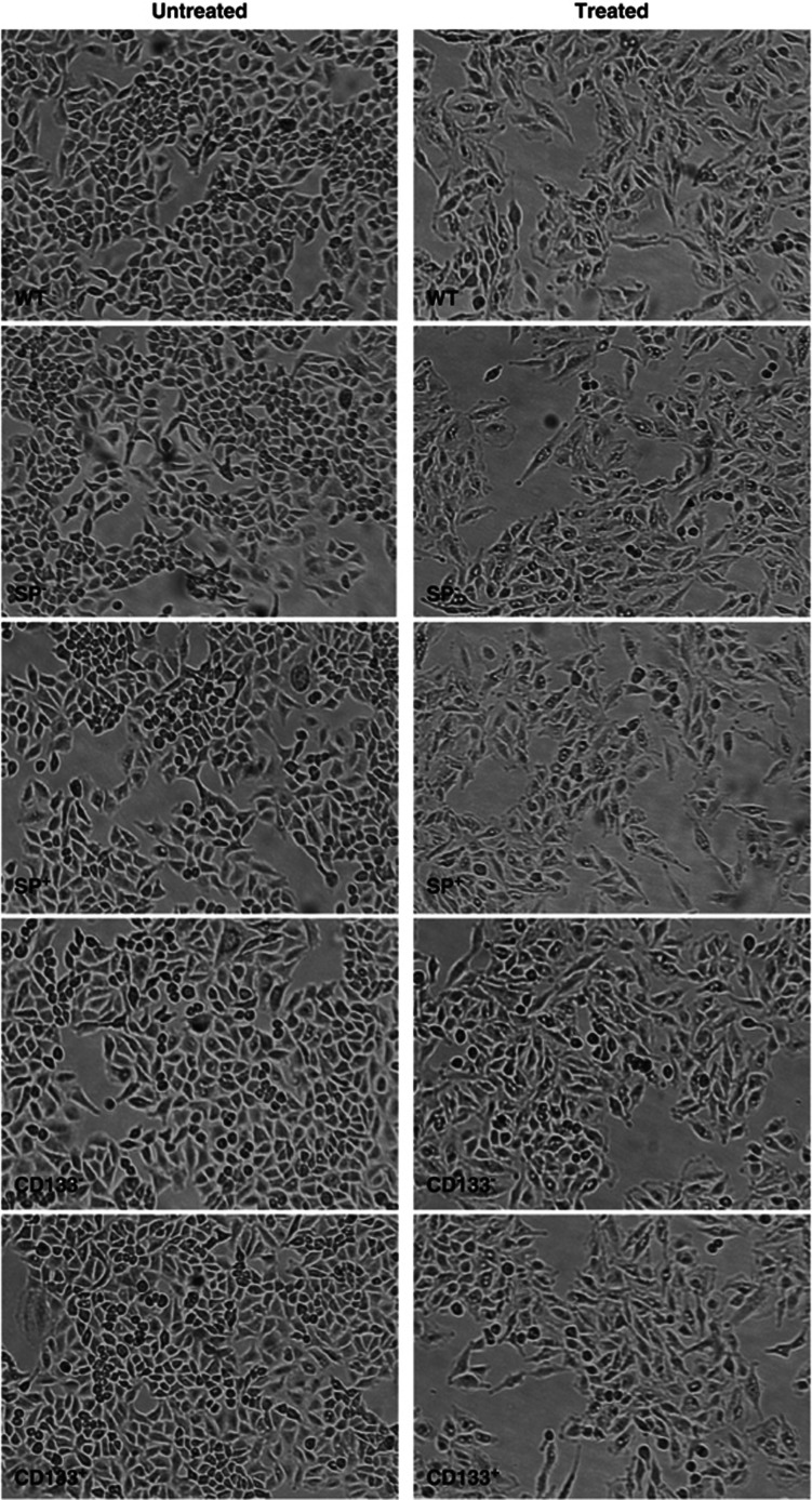
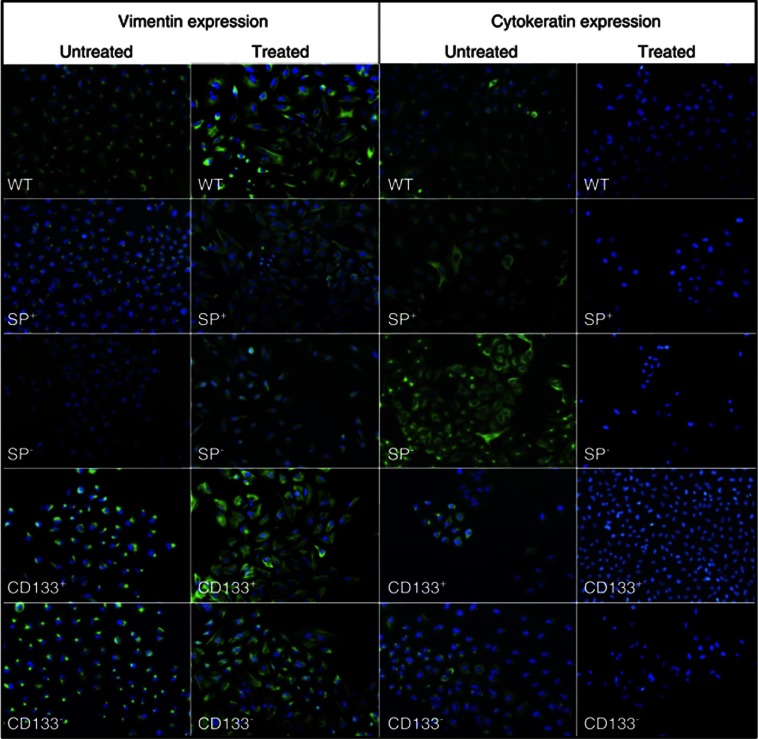
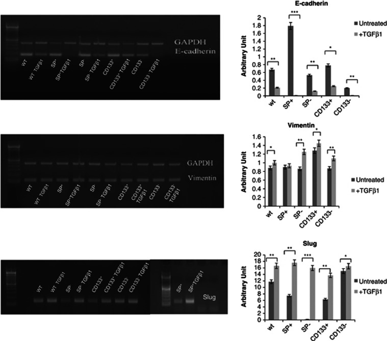
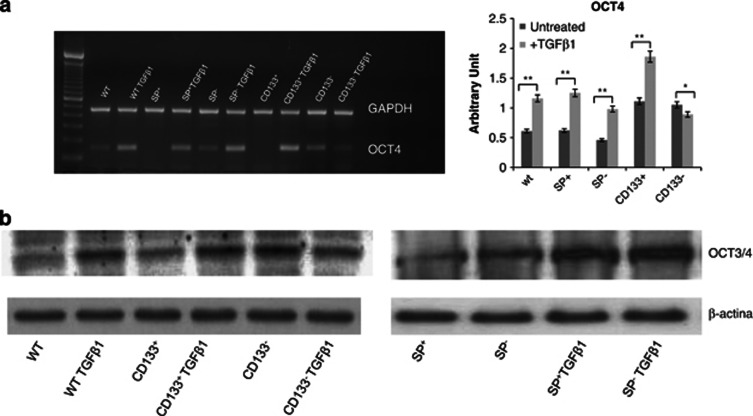
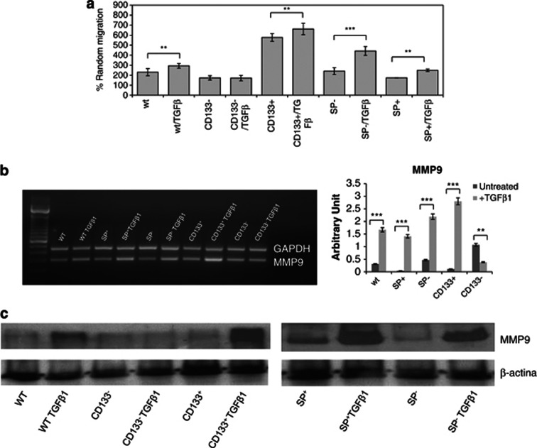
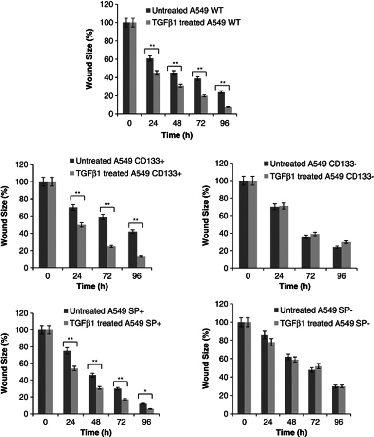
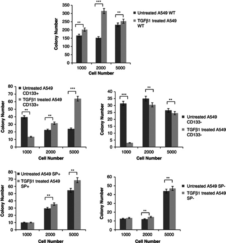
Metastasis is the leading cause of death by cancer. Non-small-cell lung cancer (NSCLC) represents nearly 85% of primary malignant lung tumours. Recent researches have demonstrated that epithelial-to-mesenchymal transition (EMT) plays a key role in the early process of metastasis of cancer cells. Transforming growth factor-β1 (TGF-β1) is the major inductor of EMT. The aim of this study is to investigate TGF-β1's effect on cancer stem cells (CSCs) identified as cells positive for CD133, side population (SP) and non-cancer stem cells (non-CSCs) identified as cells negative for CD133, and SP in the A549 cell line. We demonstrate that TGF-β1 induces EMT in both CSC and non-CSC A549 sublines, upregulating the expression of mesenchymal markers such as vimentin and Slug, and downregulating levels of epithelial markers such as e-cadherin and cytokeratins. CSC and non-CSC A549 sublines undergoing EMT show a strong migration and strong levels of MMP9 except for the CD133(-) cell fraction. OCT4 levels are strongly upregulated in all cell fractions except CD133(-) cells. On the contrary, wound size reveals that TGF-β1 enhances motility in wild-type A549 as well as CD133(+) and SP(+) cells. For CD133(-) and SP(-) cells, TGF-β1 exposure does not change the motility. Finally, assessment of growth kinetics reveals major colony-forming efficiency in CD133(+) A549 cells. In particular, SP(+) and SP(-) A549 cells show more efficiency to form colonies than untreated corresponding cells, while for CD133(-) cells no change in colony number was observable after TGF-β1 exposure. We conclude that it is possible to highlight different cell subpopulations with different grades of stemness. Each population seems to be involved in different biological mechanisms such as stemness maintenance, tumorigenicity, invasion and migration.
Figures
Similar articles
-
KLF8 involves in TGF-beta-induced EMT and promotes invasion and migration in gastric cancer cells.J Cancer Res Clin Oncol. 2013 Jun;139(6):1033-42. doi: 10.1007/s00432-012-1363-3. Epub 2013 Mar 16. J Cancer Res Clin Oncol. 2013. PMID: 23504025 Free PMC article.
-
Epithelial to mesenchymal transition by TGFβ-1 induction increases stemness characteristics in primary non small cell lung cancer cell line.PLoS One. 2011;6(6):e21548. doi: 10.1371/journal.pone.0021548. Epub 2011 Jun 30. PLoS One. 2011. PMID: 21738704 Free PMC article.
-
Norepinephrine induced epithelial-mesenchymal transition in HT-29 and A549 cells in vitro.J Cancer Res Clin Oncol. 2016 Feb;142(2):423-35. doi: 10.1007/s00432-015-2044-9. Epub 2015 Sep 10. J Cancer Res Clin Oncol. 2016. PMID: 26358081 Free PMC article.
-
A rapid and systematic review of the clinical effectiveness and cost-effectiveness of paclitaxel, docetaxel, gemcitabine and vinorelbine in non-small-cell lung cancer.Health Technol Assess. 2001;5(32):1-195. doi: 10.3310/hta5320. Health Technol Assess. 2001. PMID: 12065068
-
Impact of residual disease as a prognostic factor for survival in women with advanced epithelial ovarian cancer after primary surgery.Cochrane Database Syst Rev. 2022 Sep 26;9(9):CD015048. doi: 10.1002/14651858.CD015048.pub2. Cochrane Database Syst Rev. 2022. PMID: 36161421 Free PMC article.
Cited by
-
A peptide tag-specific nanobody enables high-quality labeling for dSTORM imaging.Nat Commun. 2018 Mar 2;9(1):930. doi: 10.1038/s41467-018-03191-2. Nat Commun. 2018. PMID: 29500346 Free PMC article.
-
CD133, Selectively Targeting the Root of Cancer.Toxins (Basel). 2016 May 28;8(6):165. doi: 10.3390/toxins8060165. Toxins (Basel). 2016. PMID: 27240402 Free PMC article. Review.
-
Utilization of lung cancer cell lines for the study of lung cancer stem cells.Oncol Lett. 2018 May;15(5):6791-6798. doi: 10.3892/ol.2018.8265. Epub 2018 Mar 14. Oncol Lett. 2018. PMID: 29731860 Free PMC article. Review.
-
CD44 silencing decreases the expression of stem cell-related factors induced by transforming growth factor β1 and tumor necrosis factor α in lung cancer: Preliminary findings.Bosn J Basic Med Sci. 2017 Aug 20;17(3):228-234. doi: 10.17305/bjbms.2017.1966. Bosn J Basic Med Sci. 2017. PMID: 28446126 Free PMC article.
-
SMAD2 linker phosphorylation impacts overall survival, proliferation, TGFβ1-dependent gene expression and pluripotency-related proteins in NSCLC.Br J Cancer. 2025 Jul;133(1):52-65. doi: 10.1038/s41416-025-02970-1. Epub 2025 May 3. Br J Cancer. 2025. PMID: 40319202 Free PMC article.
References
-
- Govindan R, Page N, Morgensztern D, Read W, Tierney R, Vlahiotis A, et al. Changing epidemiology of small-cell lung cancer in the United States over the last 30 years: analysis of the surveillance, epidemiologic, and end results database. J Clin Oncol. 2006;24:4539–4544. - PubMed
-
- World Health Organization . World Cancer Report. IARC Press: Lyon; 2008. International Agency for Research on Cancer.
-
- Jemal A, Siegel R, Xu J, Ward E. Cancer statistics, 2010. CA Cancer J Clin. 2010;60:277–300. - PubMed
-
- Gavert N, Ben-Ze'ev A. Epithelial-mesenchymal transition and the invasive potential of tumors. Trends Mol Med. 2008;14:199–209. - PubMed
-
- Nawshad A, Lagamba D, Polad A, Hay ED. Transforming growth factor-b signaling during epithelial-mesenchymal transformation: implications for embryogenesis and tumor metastasis. Cells Tissues Organs. 2005;179:11–23. - PubMed
Publication types
MeSH terms
Substances
LinkOut - more resources
Full Text Sources
Other Literature Sources
Research Materials
Miscellaneous
