. 2013 Jul;33(7):1580-90.
doi: 10.1161/ATVBAHA.112.300912. Epub 2013 May 2.

Notch1 promotes the pro-osteogenic response of human aortic valve interstitial cells via modulation of ERK1/2 and nuclear factor-κB activation

Affiliations

Notch1 promotes the pro-osteogenic response of human aortic valve interstitial cells via modulation of ERK1/2 and nuclear factor-κB activation

Qingchun Zeng et al. Arterioscler Thromb Vasc Biol. 2013 Jul.

Abstract

Objective: Calcific aortic valve disease is a leading cardiovascular disease in the elderly, and progressive calcification results in the failure of valvular function. Aortic valve interstitial cells (AVICs) from stenotic valves express higher levels of bone morphogenetic protein-2 in response to Toll-like receptor 4 stimulation. We recently found that Toll-like receptor 4 interacts with Notch1 in human AVICs. This study tests the hypothesis that Notch1 promotes the pro-osteogenic response of human AVICs.

Approach and results: AVICs isolated from diseased human valves expressed higher levels of bone morphogenetic protein-2 and alkaline phosphatase after lipopolysaccharide stimulation. The augmented pro-osteogenic response is associated with elevated cellular levels of Notch1 and enhanced Notch1 cleavage in response to lipopolysaccharide stimulation. Inhibition or silencing of Notch1 suppressed the pro-osteogenic response in diseased cells, and the Notch 1 ligand, Jagged1, enhanced the response in AVICs isolated from normal human valves. Interestingly, extracellular signal-regulated protein kinases 1/2 (ERK1/2) and nuclear factor-κB phosphorylation induced by lipopolysaccharide was markedly reduced by inhibition or silencing of Notch1 and enhanced by Jagged1. Inhibition of ERK1/2 or nuclear factor-κB also reduced bone morphogenetic protein-2 and alkaline phosphatase expression induced by lipopolysaccharide.

Conclusions: Notch1 mediates the pro-osteogenic response to Toll-like receptor 4 stimulation in human AVICs. Elevated Notch1 levels and enhanced Notch1 activation play a major role in augmentation of the pro-osteogenic response of AVICs of stenotic valves through modulation of ERK1/2 and nuclear factor-κB activation. These pathways could be potential therapeutic targets for prevention of the progression of calcific aortic valve disease.

Keywords: Notch1; Toll-like receptor 4; aortic valve; pro-osteogenic proteins; signal transduction.


Figures

Figure 1
Figure 1. Exaggerated pro-osteogenic response to TLR4 stimulation in AVICs of stenotic valves is associated with elevated Notch1 levels and enhanced Notch1 cleavage
A–D. AVICs of normal and stenotic valves are treated with LPS (200 ng/ml) for 1–28 days. A. Representative immunoblots, densitometric data and ELISA data (from 5 separate experiments using different cell isolates) show that cells from stenotic valves produce higher levels of BMP-2 and release greater amounts of this osteogenic factor after LPS stimulation for 24 h. B. Representative immunoblots, densitometric data and enzyme activity images (from 5 separate experiments using different cell isolates) show that cells from stenotic valves express higher levels of ALP at 24 h and exhibit greater ALP activity at 14 days of LPS stimulation. C. Representative images of 3 separate experiments and quantitative data show greater formation of calcium deposits (arrow) in AVICs from stenotic valves following LPS stimulation for 4 weeks. D. Representative images of aortic valve tissue show increased number of cells with Notch1 immunoreactivity in stenotic valves and higher density of Notch1 immunohistochemical staining in the interstitial cells of stenotic valves (20× objective). Immunoblots and densitometric data show that AVICs from 5 stenotic valves have higher levels of Notch1 protein. E. Representative immunoblots and densitometric data (from 5 separate experiments using different cell isolates) show that TLR4 stimulation induces Notch1 cleavage in AVICs of normal and stenotic valves. Cells of stenotic valves generate higher levels of NICD1. F. Representative images of immunofluorescense staining show that TLR4 stimulation induces Notch1 (Cy3) translocation from the cell surface (FITC) to perinuclear regions and into the nuclei (DAPI) in AVICs of stenotic valves. *P<0.05 vs. corresponding control; †P<0.05 vs. normal cells receiving the same treatment.
Figure 2
Figure 2. Notch1 plays an important role in mediating the pro-osteogenic response in AVICs of stenotic valves
A. AVICs of normal and stenotic aortic valves are stimulated with LPS in the presence or absence of γ-secretase inhibitor DAPT (50 μM). Representative immunoblots and densitometric data (from 5 separate experiments using different cell isolates) show that DAPT reduces the levels of BMP-2 and ALP in AVICs of normal and stenotic valves at 24 h of LPS treatment. The effect of Notch1 inhibition is greater in AVICs of stenotic valves. Representative images of 3 separate experiments and quantitative data show that inhibition of Notch1 with DAPT reduces calcium deposits (arrow) in AVICs of stenotic valves following LPS treatment for 4 weeks. B. Representative immunoblots and densitometric data (from 5 separate experiments using different cell isolates) show that AVICs of stenotic aortic valves treated with Notch1 siRNA have markedly lower levels of Notch1 protein and attenuated BMP-2 and ALP expression following TLR4 stimulation with LPS for 24 h. C. Representative immunoblots and densitometric data (from 5 separate experiments using different cell isolates) show that Notch1 ligand Jagged1 enhances Notch1 cleavage in AVICs of normal valves and up-regulates BMP-2 and ALP expression after treatment with LPS for 24 h. *P<0.05 vs. corresponding control; †P<0.05 vs. normal cells receiving the same treatment; ‡P<0.05 vs. LPS alone.
Figure 3
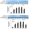
Figure 3. The exaggerated pro-osteogenic response in AVICs of stenotic valves correlates with enhanced activation of ERK1/2 and NF-κB
Representative immunoblots and densitometric data (from 5 separate experiments using different cell isolates) show that AVICs of stenotic valves exhibit enhanced ERK1/2 and NF-κB phosphorylation after treatment with LPS for 1–24 h. *P<0.05 vs. control; †P<0.05 vs. normal cells receiving the same treatment.
Figure 4
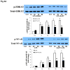
Figure 4. Notch1 modulates ERK1/2 and NF-κB phosphorylation
A. AVICs of stenotic valves are treated with DAPT and then stimulated with LPS for 1–8 h. Representative immunoblots and densitometric data (n=5) show that inhibition of Notch1 with DAPT markedly reduces ERK1/2 and NF-κB p65 phosphorylation. B. AVICs of stenotic aortic valves are treated with Notch1 siRNA and then stimulated with LPS for 1–4 h. Representative immunoblots and densitometric data (from 5 separate experiments using different cell isolates) show that Notch1 knockdown reduces ERK1/2 and NF-κB p65 phosphorylation. C. AVICs of normal valves are cultured on Jagged1-coated plates and stimulated with LPS for 1–8 h. Representative immunoblots and densitometric data (from 5 separate experiments using different cell isolates) show that Notch1 ligand Jagged1 enhances ERK1/2 and NF-κB p65 phosphorylation. D. AVICs of normal valves were cultured on Jagged1- coated plates and then treated with LPS for 1–8 h. A representative immunoblot of 3 separate experiments shows that activation of Notch1 with Jagged1 enhances MEK1/2 phosphorylation in AVICs exposed to LPS. E. AVICs of stenotic valves were treated with PD98059 and then stimulated with LPS for 1–8 h. Representative immunoblots of 5 separate experiments show that PD98059 suppresses ERK1/2 phosphorylation, and inhibition of the ERK1/2 pathway with PD98059 attenuates NF-κB p65 phosphorylation. *P<0.05 vs. corresponding control; ‡P<0.05 vs. LPS alone or vehicle+LPS.
Figure 5
Figure 5. The ERK1/2 and NF-κB pathways mediate BMP-2 and ALP expression in AVICs of stenotic valves
A. AVICs of stenotic aortic valves are treated with PD98059 (25 μM) 1 h prior to treatment with LPS. Representative immunoblots and densitometric data (from 5 separate experiments using different cell isolates) show that inhibition of the ERK1/2 pathway attenuates BMP-2 and ALP expression following LPS treatment for 24 h. Representative images of 3 separate and quantitative data show that inhibition of the ERK1/2 pathway with PD98059 reduces calcium deposits (arrow) in AVICs of stenotic valves following LPS treatment for 4 weeks. B. AVICs of stenotic aortic valves are treated with a cell-permeable NF-κB inhibitory peptide (SN50, 100 μg/ml; the same concentration of SN50M for control) 1 h prior to treatment with LPS. Representative immunoblots and densitometric data (from 5 separate experiments using different cell isolates) show that inhibition of NF-κB reduces BMP-2 and ALP expression following LPS treatment for 24 h. Representative images of 3 separate experiments and quantitative data show that inhibition of NF-κB reduces calcium deposits (arrow) in AVICs of stenotic valves following LPS treatment for 4 weeks. *P<0.05 vs. control, ‡P<0.05 vs. LPS alone or SN50M+LPS.
Figure 6
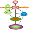
Figure 6. Schematic diagram of the up-regulation of the pro-osteogenic response by Notch1
In AVICs of stenotic valves, stimulation of TLR4 with LPS induces enhanced activation of Notch1, and augmented phosphorylation of ERK1/2 and NF-κB. Notch1 modulates ERK1/2 and NF-κB activity through its intracellular domain NICD1. Both the ERK1/2 and NF-κB pathways are involved in up-regulating the expression of pro-osteogenic proteins. It appears that the ERK1/2 pathway up-regulates the pro-osteogenic response in human AVICs primarily through modulation of NF-κB activation.

Similar articles

Cited by

References

    1. Mohler ER, 3rd, Gannon F, Reynolds C, Zimmerman R, Keane MG, Kaplan FS. Bone formation and inflammation in cardiac valves. Circulation. 2001;103:1522–1528. - PubMed
    1. Rajamannan NM, Gersh B, Bonow RO. Calcific aortic stenosis: from bench to the bedside--emerging clinical and cellular concepts. Heart. 2003;89:801–805. - PMC - PubMed
    1. Rajamannan NM, Evans FJ, Aikawa E, Grande-Allen KJ, Demer LL, Heistad DD, Simmons CA, Masters KS, Mathieu P, O’Brien KD, Schoen FJ, Towler DA, Yoganathan AP, Otto CM. Calcific aortic valve disease: not simply a degenerative process: A review and agenda for research from the National Heart and Lung and Blood Institute Aortic Stenosis Working Group. Executive summary: Calcific aortic valve disease-2011 update. Circulation. 2011;124:1783–1791. - PMC - PubMed
    1. New SE, Aikawa E. Molecular imaging insights into early inflammatory stages of arterial and aortic valve calcification. Circ Res. 2011;108:1381–1391. - PMC - PubMed
    1. Osman L, Yacoub MH, Latif N, Amrani M, Chester AH. Role of human valve interstitial cells in valve calcification and their response to atorvastatin. Circulation. 2006;114:I547–552. - PubMed

Publication types

MeSH terms

Substances

Supplementary concepts