. 2013 Jul;33(7):1544-51.
doi: 10.1161/ATVBAHA.112.301165. Epub 2013 May 2.

p21 activated kinase signaling coordinates glycoprotein receptor VI-mediated platelet aggregation, lamellipodia formation, and aggregate stability under shear

Affiliations

p21 activated kinase signaling coordinates glycoprotein receptor VI-mediated platelet aggregation, lamellipodia formation, and aggregate stability under shear

Joseph E Aslan et al. Arterioscler Thromb Vasc Biol. 2013 Jul.

Abstract

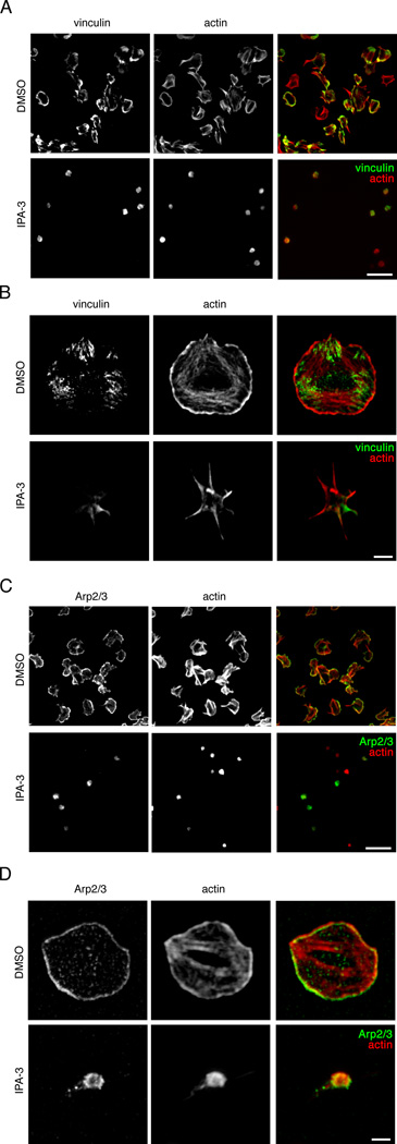
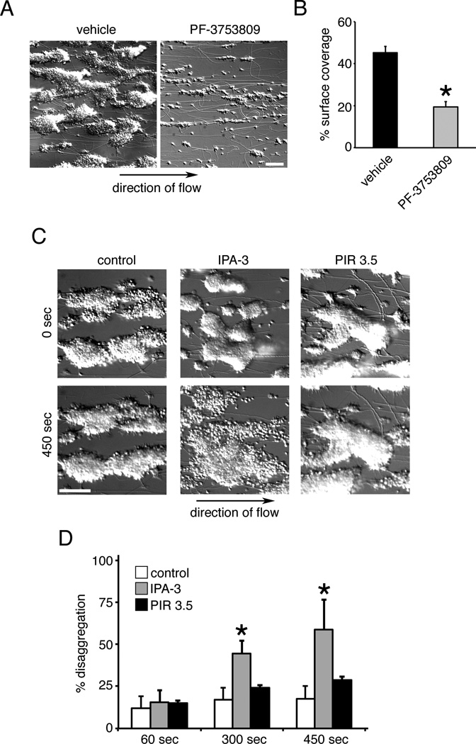
Objective: Rho GTPase proteins play a central role in regulating the dynamics of the platelet actin cytoskeleton. Yet, little is known regarding how Rho GTPase activation coordinates platelet activation and function. In this study, we aimed to characterize the role of the Rho GTPase effector, p21 activated kinase (PAK), in platelet activation, lamellipodia formation, and aggregate formation under shear.

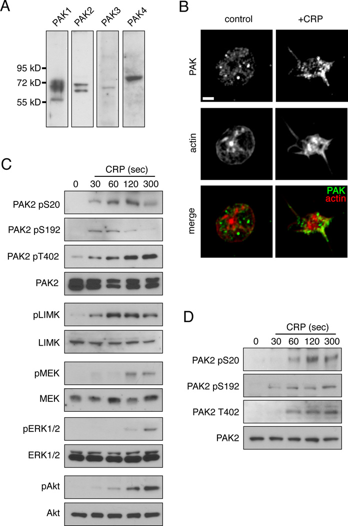
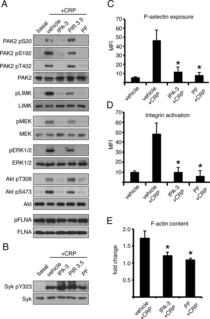
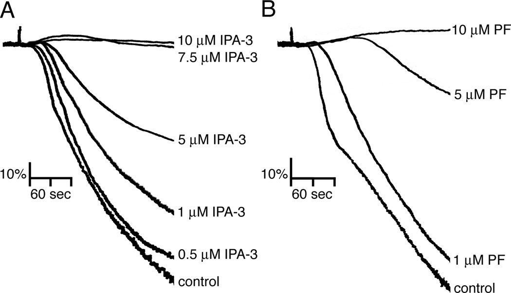
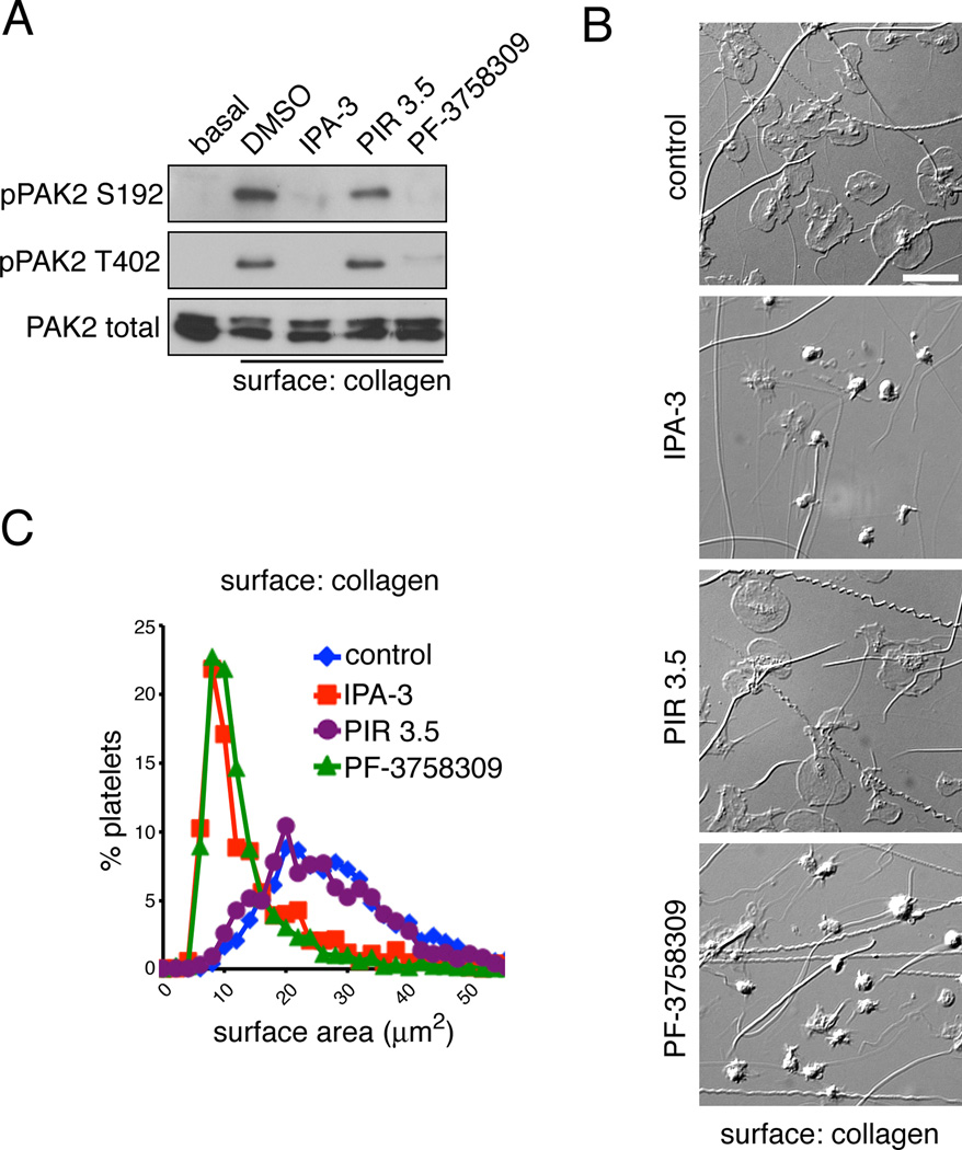
Approach and results: Stimulation of platelets with the glycoprotein receptor VI agonist, collagen-related peptide, rapidly activated PAK in a time course preceding phosphorylation of PAK substrates, LIM domain kinase LIMK1 and the MAPK/ERK kinase MEK, and the subsequent activation of MAPKs and Akt. Pharmacological inhibitors of PAK blocked signaling events downstream of PAK and prevented platelet secretion as well as platelet aggregation in response to collagen-related peptide. PAK inhibitors also prevented PAK activation and platelet spreading on collagen surfaces. PAK was also required for the formation of platelet aggregates and to maintain aggregate stability under physiological shear flow conditions.

Conclusions: These results suggest that PAK serves as an orchestrator of platelet functional responses after activation downstream of the platelet collagen receptor, glycoprotein receptor VI.

Keywords: cell signaling; platelet; signal transduction.


Figures

Figure 1
Figure 1. Platelet treatment with CRP stimulates PAK activation and the phosphorylation of PAK effectors
(A) Replicate samples of human platelet lysates (50 µg total protein) were assayed for PAK isoform expression by Western blot. (B) Super resolution fluorescent microscopy analysis of PAK localization. Replicate samples of control and CRP stimulated human platelets were fixed and stained for PAK and actin. Scale bar = 1 µm. (C) Time course of platelet PAK phosphorylation and activation upon stimulation with CRP. Replicate samples of purified human platelets (5 × 10⁸/ml) were treated with CRP (1 µg/ml) for 30, 60, 120 and 300 sec before lysis and Western blot analysis for PAK, LIMK, MEK, ERK and Akt phosphorylation. (D) Time course of platelet PAK phosphorylation in response to CRP stimulation in the presence of apyrase (2 U/ml), indomethacin (10 µM) and eptifibatide (20 µg/ml).
Figure 2
Figure 2. PAK activity is required for platelet PAK effector phosphorylation, secretion, integrin activation and F-actin formation
(A) Replicate samples of purified human platelets (5 × 10⁸/ml) treated with vehicle (0.1% DMSO), IPA-3 (10 µM), PIR 3.5 (10 µM) or PF-3758309 (10 µM) for 10 min prior to stimulation with CRP (1 µg/ml, 300 sec) in the presence of apyrase (2 U/ml) and eptifibatide (20 µg/ml). After lysis, samples were analyzed for PAK2 Ser20, Ser192 and Thr402 autosphosphorylation, the phosphorylation PAK effectors LIMK1 (Thr508) and MEK1/2 (Ser217/221) and the activation of ERK and Akt signaling. Results representative of four experiments are shown. (B) Platelets were also examined for Src-mediated phosphorylation of Syk Tyr323. (C) Platelet P-selectin, (D) integrin αIIbβ3 activation and (E) F-actin content analyzed by flow cytometry following vehicle (DMSO), IPA-3 and PF-3758309 treatment and CRP stimulation. Data are represented as mean ± SEM. Significant results (p < 0.05) are indicated with an asterisk.
Figure 3
Figure 3. Inhibition of PAK blocks platelet aggregation in response to CRP
Washed human platelets (2 × 10⁸/ml) were incubated with vehicle (DMSO) or increasing concentrations of (A) IPA-3 or (B) PF-123758309 in the presence of 2 U/ml apyrase prior to stimulation with CRP (1 µg/ml) and the change in optical density indicative of platelet aggregation was recorded. Representative aggregation traces of four separate experiments are shown.
Figure 4
Figure 4. PAK activation is required for spreading on a surface of collagen
(A) Replicate samples of purified human platelets (5 × 10⁸/ml) were treated with vehicle (DMSO), IPA-3 (10 µM), PIR 3.5 (10 µM) or PF-3758309 (10 µM) for 10 min prior to exposure to a collagen surface in the presence of 2 U/ml apyrase. After 45 min, platelets were stimulated lysed and analyzed for PAK activation by Western blot for PAK2-pSer192 and PAK2-pThr402. (B) Replicate wells of collagen-coated coverglass were seeded with human platelets (2 × 10⁷/ml) for 45 min after pretreatment with vehicle (DMSO), IPA-3 (10 µM), PIR 3.5 (10 µM) or PF-3758309 (10 µM). Platelets were fixed and analyzed by DIC microscopy. Images representative of five experiments are shown. Scale bar = 10 µm. (C) Quantification and distribution of platelet surface areas (n = 200 per condition) under control and PAK-inhibited conditions.
Figure 5
Figure 5. PAK activity regulates actin-rich adhesion and lamellipodia formation in platelets spread on collagen
Replicate wells of collagen-coated coverglass were seeded with human platelets (2 × 10⁷/ml) for 45 min after pretreatment with vehicle (DMSO), IPA-3 (10 µM), PIR 3.5 (10 µM) or PF-3758309 (10 µM). Platelets were fixed, stained for (A,B) vinculin and actin or (C,D) Arp2/3 and actin and analyzed by fluorescence microscopy. Images representative of five experiments are shown. (A,C) Scale bar = 10 µm. Super resolution microscopy analysis of (B) platelet focal adhesion formation and (D) lamellipodia formation of vehicle (DMSO) and IPA-3 (10 µM) treated platelets on a surface of collagen, visualized by SR-SIM of actin (red) and vinculin or Arp2/3 (green) staining. Scale bar = 2 µm.
Figure 6
Figure 6. Platelet aggregate formation and stability under shear requires PAK
(A) Human whole blood was treated with vehicle or PF-3758309 (20 µM) and perfused over collagen at a shear rate of 1500 s-1 to produce platelet aggregates. (B) Quantification of percentage of surface area coverage by platelet aggregates in the presence of vehicle or PF-3758309 (n = 3). (C) Platelet aggregates were formed following perfusion of PPACK-anticoagulated blood over collagen for 4 min at a wall shear rate of 1500 s-1. Following washing for 4 min with buffer containing fibrinogen (3 mg/ml), platelet aggregates were perfused with buffer containing fibrinogen and vehicle (DMSO), IPA-3 (10 µM) or PIR 3.5 (10 µM). Representative DIC images are shown. Scale bar = 100 µm. (D) Quantification of platelet disaggregation promoted by IPA-3 under flow (n = 3). Data are represented as mean ± SEM. Significant results (p < 0.05) are indicated with an asterisk.

References

    1. Furie B, Furie BC. Thrombus formation in vivo. J Clin Invest. 2005;115:3355–3362. - PMC - PubMed
    1. Jackson SP, Schoenwaelder SM. Antiplatelet therapy: in search of the 'magic bullet'. Nat Rev Drug Discov. 2003;2:775–789. - PubMed
    1. Watson SP, Auger JM, McCarty OJ, Pearce AC. GPVI and integrin alphaIIb beta3 signaling in platelets. J Thromb Haemost. 2005;3:1752–1762. - PubMed
    1. Calaminus SD, McCarty OJ, Auger JM, Pearce AC, Insall RH, Watson SP, Machesky LM. A major role for Scar/WAVE-1 downstream of GPVI in platelets. J Thromb Haemost. 2007;5:535–541. - PMC - PubMed
    1. Li Z, Kim ES, Bearer EL. Arp2/3 complex is required for actin polymerization during platelet shape change. Blood. 2002;99:4466–4474. - PMC - PubMed

Publication types

MeSH terms