Review
. 2013 Mar;137(3):455-93.

Tuberculosis: From an incurable scourge to a curable disease - journey over a millennium

Affiliations
Review

Tuberculosis: From an incurable scourge to a curable disease - journey over a millennium

Surendra K Sharma et al. Indian J Med Res. 2013 Mar.

Abstract

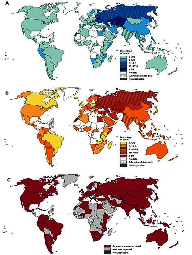
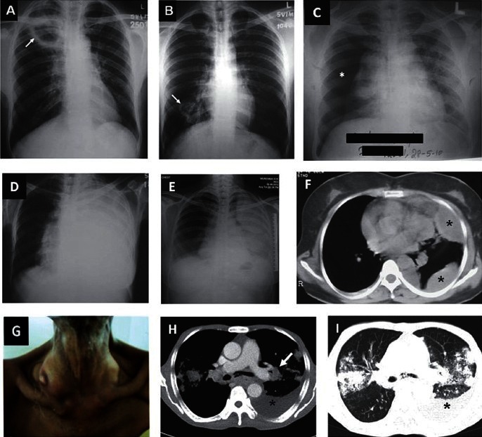
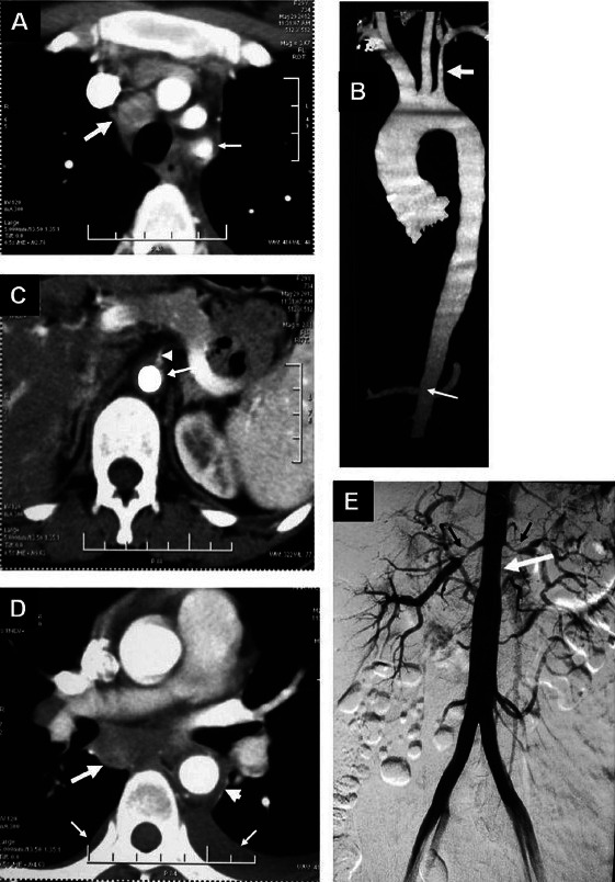
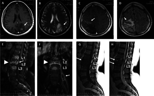
Globally, tuberculosis (TB) still remains a major public health problem. India is a high TB burden country contributing to 26 per cent of global TB burden. During 1944-1980, TB became treatable and short-course chemotherapy emerged as the standard of care. When TB elimination seemed possible in the early 1980s, global human immunodeficiency virus (HIV) infection/acquired immunodeficiency syndrome (AIDS) pandemic resulted in a resurgence of TB. Widespread occurrence of multidrug-resistant and extensively drug-resistant TB (M/XDR-TB) is threatening to destabilize TB control globally. Atypical clinical presentation still poses a challenge. Disseminated, miliary and cryptic TB are being increasingly recognized. Availability of newer imaging modalities has allowed more efficient localization of lesions and use of image guided procedures has facilitated definitive diagnosis of extrapulmonary TB. Introduction of liquid culture, rapid drug-susceptibility testing (DST), molecular diagnostic methods has helped in rapid detection, speciation and DST profiling of Mycobacterium tuberculosis isolates. While treatment of TB and HIV-TB co-infection has become simpler, efforts are on to shorten the treatment duration. However, drug toxicities and drug-drug interactions still constitute a significant challenge. Recently, there has been better understanding of anti-TB drug-induced hepatotoxicity and its frequent confounding by viral hepatitis, especially, in resource-constrained settings; and immune reconstitution inflammatory syndrome (IRIS) in HIV-TB. Quest for newer biomarkers for predicting a durable cure, relapse, discovery/repurposing of newer anti-TB drugs, development of newer vaccines continues to achieve the goal of eliminating TB altogether by 2050.


Figures

Fig. 1
Fig. 1
(A). Distribution of percentage of new tuberculosis cases with MDR-TB 1994-2011. (B). Distribution of percentage of previously treated tuberculosis cases with MDR-TB 1994-2011. (C). Countries that had reported at least one XDR-TB case 1994-2011. MDR-TB, multidrug-resistant tuberculosis; XDR-TB, extensively drug-resistant tuberculosis. Reproduced with permission from World Health Organization (reference 41)
Fig. 2
Fig. 2
Natural history of Mycobacterium tuberculosis infection and scope for intervention. TB, tuberculosis; BCG, bacille Calmette-Guerin; HIV, human immunodeficiency virus; AIDS, acquired immunodeficiency syndrome; TNF, tumour necrosis factor; LTBI, latent TB infection; SS, sputum smear. Adapted and updated from references , , .
Fig. 3
Fig. 3
(A) Chest radiograph (postero-anterior view) showing a cavity in the right upper zone (arrow), and (B) lower zone (arrow). (C). Chest radiograph (postero-anterior view) in a patient presenting to the emergency room with severe breathlessness showing right-sided pneumothorax (asterisk). Sputum smear was positive for acid-fast bacilli. (D). Chest radiograph (poster-anterior view) showing left-sided massive pleural effusion. (E). Chest radiograph (poster-anterior view) in another patient showing left-sided loculated pleural effusion. (F). CECT (chest) of the same patient showing left-sided loculated pleural effusion (asterisks). (G) Clinical photograph of a patient with disseminated TB showing right-sided cervical lymphadenopathy with cold abscess. (H) CECT chest of the same patient showing left-sided pleural effusion (asterisk), left hilar lymphadenopathy [arrow (mediastinal window)]; and (I) and bilateral parenchymal infiltrates and left sided pleural effusion (asterisk) (lung window).
Fig. 4
Fig. 4
Nonspecific aortoarteritis. (A) Axial contrast enhanced CT showing an enlarged mediastinal lymph node (thick arrow) and diffuse wall thickening of left subclavian artery (Thin arrow) (B). Volume rendered CT angiography image showing diffuse long segment narrowing of left subclavian artery (thick arrow) and abdominal aorta at the renal artery origin level as well as infrarenal segment (thin arrow) (C). Axial contrast enhanced CT showing diffuse wall thickening of abdominal aorta (long arrow) with luminal narrowing of the origin of superior mesenteric artery (arrow head) (D). Axial contrast enhanced CT showing an enlarged subcarinal mediastinal lymph node (thick arrow) and diffuse wall thickening of descending thoracic aorta (arrow head). Bilateral pleural effusions [left more than right (thin arrows)] can also be seen (E). Digital substraction angiography (DSA) showing diffuse long segment narrowing of abdominal aorta at the renal artery origin level and infrarenal segment (white arrow). There is a marked narrowing of the bilateral renal arteries (black arrows)
Fig. 5
Fig. 5
(A) T1-weighted pre-contrast and (B) contrast-enhanced MRI images showing ring enhancing lesion with perileional oedema in the left parietal lobe (arrows). (C) T1-weighted axial MRI image showing conglomerate ring enhancing lesions in the right frontoparietal regions (arrow) (D). FLAIR sequence showing hypointense lesions with perilesional oedema (arrow) (E). T1-weighted contrast enhanced MRI showing collapse of L2 vertebral body with abnormal enhancement in L1-L3 vertebrae, prevertebral regions (arrow-head) and epidural abscess (arrow) at L2 (F). Coronal image of the same patient showing collapse of L2 vertebral body (arrow-head) and left-sided psoas abscess (arrow). (G) Pre-Contrast T1-weighted sagittal MRI showing hypointense signal in L1-L3 vertebral bodies (arrow). T2-weighted sagittal image showing hyperintensities in the end-plates of L1-L2 and L2-L3 vertebral bodies and intervening discs (arrows).
Fig. 6
Fig. 6
Intrathoracic lymph node TB. (A) CT of the chest (mediastinal window), FDG-PET (B) showing left-sided hilar lymphadenopathy (arrow). (C) PET-CT image of the same patient showing increased uptake in the lesion (arrow). (D,E,F, upper panel). Nonspecific aortoarteritis. PET-CT images showing increased uptake (arrows) at the time of initial presentation. (G,H,I lower panel) The post-treatment images of the same patient shows significant decrease in the uptake suggestive of resolution of lesions with treatment. Kind courtesy: Dr TC Kalawat, Department of Nuclear Medicine, Sri Venkateswara Institute of Medical Sciences, Tirupati (Figures 6 A-C and Drs. Arun Malhotra, Rakesh Kumar, Department of Nuclear Medicine, All India Institute of Medical Sciences, New Delhi (Figures 6D-I)
Fig. 7
Fig. 7
(A) 99mTc-methylene diphosphonte (MDP) whole body anterior and (B) posterior sweep views in a patient presenting with backache and low-grade fever showing diffuse increased radiotracer localization in the body of L4 and 5 vertebrae (arrows) suggestive of spinal TB. (Kind courtesy: Dr TC Kalawat, Department of Nuclear Medicine, Sri Venkateswara Institute of Medical Sciences, Tirupati)
Fig. 8
Fig. 8
A brief history of development of antituberculosis drugs.
Fig. 9
Fig. 9
Guidelines on timing of antiretroviral treatment in patients with HIV-TB co-infection ART, antiretroviral treatment; BHIVA, British HIV Association; EFV, efavirenz; HAART, highly active antiretroviral treatment; HIV, human immunodeficiency virus; NNRTI, non-nucleoside reverse transcriptase inhibitors; Source: References ,
Box 1
Box 1
Key issues in monitoring treatment of TB
Box 2
Box 2
Latent TB infection: key issues
Box 3
Box 3
Global efforts at TB control (1991-2013): a journey

Similar articles

Cited by

References

    1. Mohan A, Sharma SK. History. In: Sharma SK, Mohan A, editors. Tuberculosis. 2nd ed. New Delhi: Jaypee Brothers Medical Publishers; 2009. pp. 7–15.
    1. Rubin SA. Tuberculosis. Captain of all these men of death. Radiol Clin North Am. 1995;33:619–39. - PubMed
    1. Rosenblatt MB. Pulmonary tuberculosis: evolution of modern therapy. Bull NY Acad Med. 1973;49:163–96. - PMC - PubMed
    1. Dubos R, Dubos J. Boston: Little, Brown and Company; 1952. The white plague. Tuberculosis, man and society.
    1. Waksman SA. Berkeley and Los Angeles: University of California Press; 1964. The conquest of tuberculosis.

MeSH terms

Substances