

doi: 10.1007/s12471-013-0426-7.
A calcified sinutubular junction: the discovery of a supravalvular aortic stenosis in an elderly woman
Affiliations
- PMID: 23640577
- PMCID: PMC3833916
- DOI: 10.1007/s12471-013-0426-7
Item in Clipboard
A calcified sinutubular junction: the discovery of a supravalvular aortic stenosis in an elderly woman
Neth Heart J.
2013 Dec.
Abstract
We report a case of a 64 year old woman with a calcified ring at the level of the sinotubular junction. Echocardiography and Computed Tomography showed a supravalvular aortic stenosis, without known associated lesions, except for the existence of an aberrant right subclavian artery. These combination of abnormalities makes it an unique case. Differential diagnosis of sinutubular calcification is added. From the literature a short review of supravalvular aortic stenosis is presented with indications for surgical intervention. Lifelong and regular follow up is necessary.
Figures
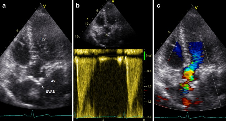
Cardiac echocardiography with apical views demonstrating the SVAS (a, left, arrow). Pulsed-wave Doppler measurement shows a maximal velocity of 1.8 m/s across the stenosis (b, middle). Colour Doppler visualises the turbulence at the level of the ST junction (c, right)
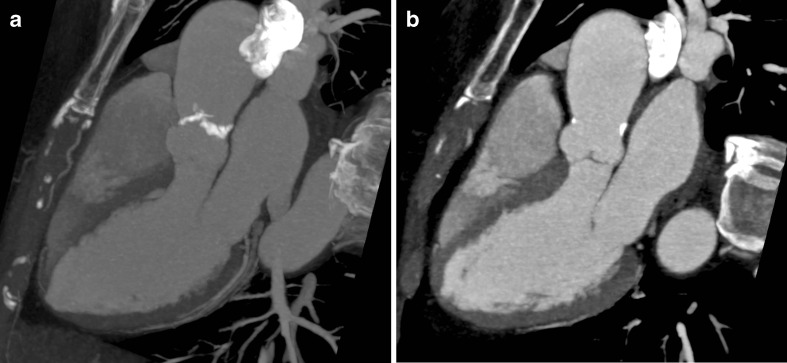
CT images in a three-chamber plane. Thick (a, left) and thin (b, right) slice maximum intensity projection (MIP) reconstructions, visualising the calcified ST ridge

CT MIP reconstructions. Axial-oblique plane (a, left) showing the calcified ring (arrow, SVAS) and axial plane (b, right) showing the aberrant right subclavian artery (arrow, LUSORIA) originating from the distal arcus aortae (arrow, ARCUS)
Similar articles
-
Patch annulo-aortoplasty in an adult patient with congenital supravalvular aortic stenosis and a small aortic annulus.Gen Thorac Cardiovasc Surg. 2011 Aug;59(8):569-71. doi: 10.1007/s11748-010-0736-2. Epub 2011 Aug 18. Gen Thorac Cardiovasc Surg. 2011. PMID: 21850585
-
[Supravalvular aortic stenosis. Report of 3 cases].Ital Heart J Suppl. 2001 Mar;2(3):307-11. Ital Heart J Suppl. 2001. PMID: 11307789 Italian.
-
Supravalvular aortic stenosis: repair with the Yacoub procedure.J Heart Valve Dis. 2004 Nov;13(6):921-4. J Heart Valve Dis. 2004. PMID: 15597582
-
Congenital supravalvar aortic stenosis: a simple lesion?Eur J Cardiothorac Surg. 2001 Feb;19(2):195-202. doi: 10.1016/s1010-7940(00)00647-3. Eur J Cardiothorac Surg. 2001. PMID: 11167112 Review.
-
Supravalvular aortic stenosis in the adult. A case presentation with unique associated features.S Afr Med J. 1981 May 23;59(22):796-803. S Afr Med J. 1981. PMID: 7015531 Review.
References
-
- Liu CW, Hwang B, Lee BC, et al. Aortic stenosis in children: 19-year experience. Zhonghua Yi Xue Za Zhi. 1997;59:107–13. - PubMed
LinkOut - more resources
Full Text Sources
Other Literature Sources
