. 2013 Jun 21;288(25):18146-61.
doi: 10.1074/jbc.M112.436584. Epub 2013 May 2.

Lacritin rescues stressed epithelia via rapid forkhead box O3 (FOXO3)-associated autophagy that restores metabolism

Affiliations

Lacritin rescues stressed epithelia via rapid forkhead box O3 (FOXO3)-associated autophagy that restores metabolism

Ningning Wang et al. J Biol Chem. .

Abstract

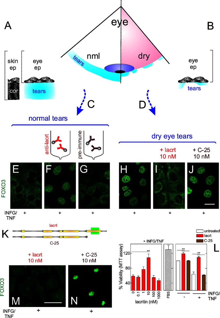
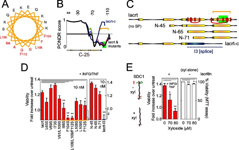
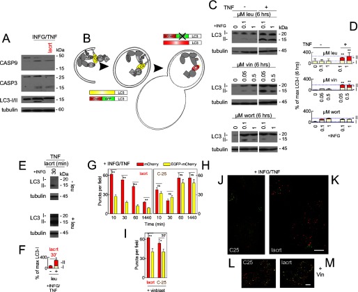
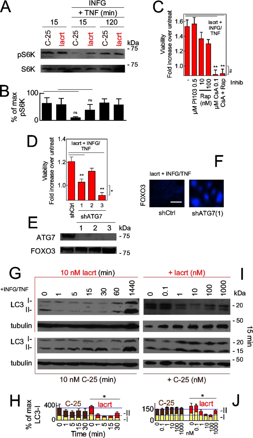
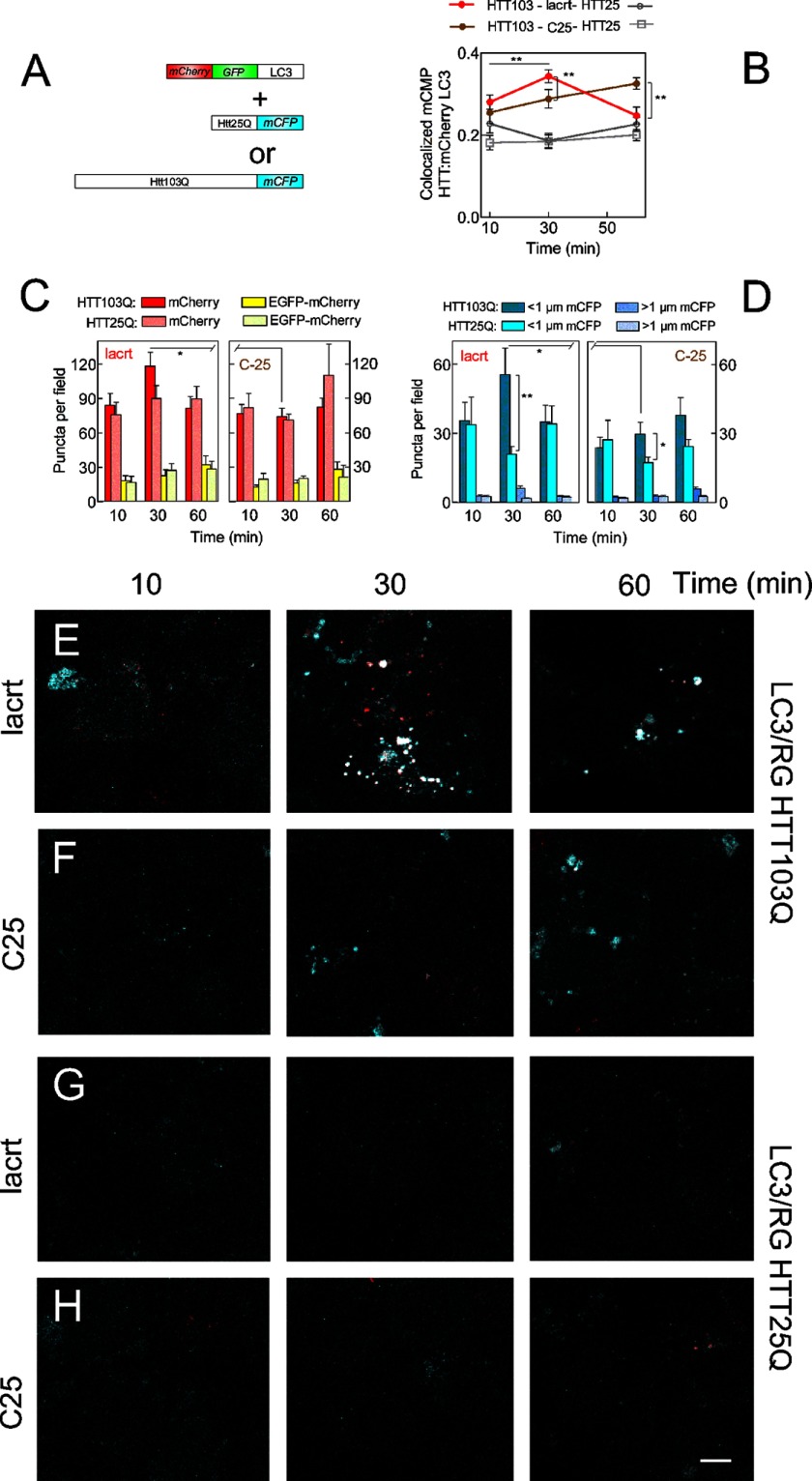
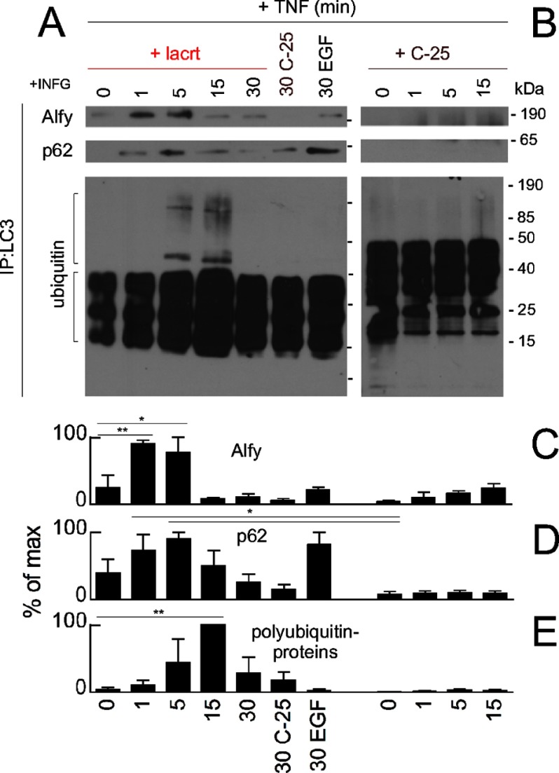
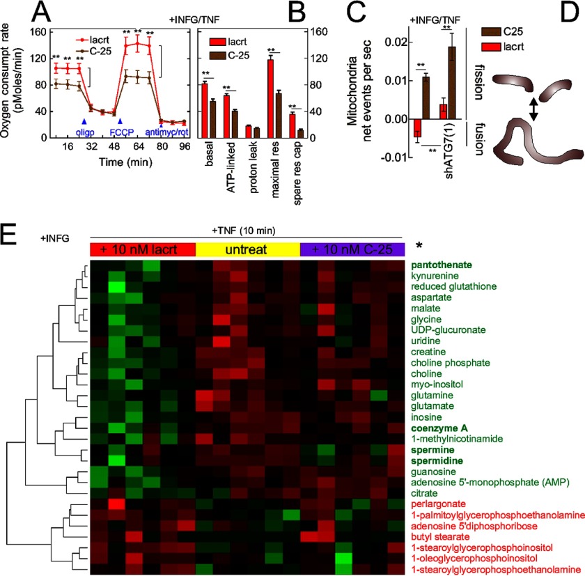
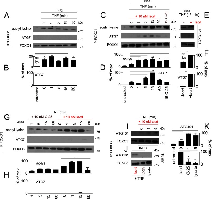
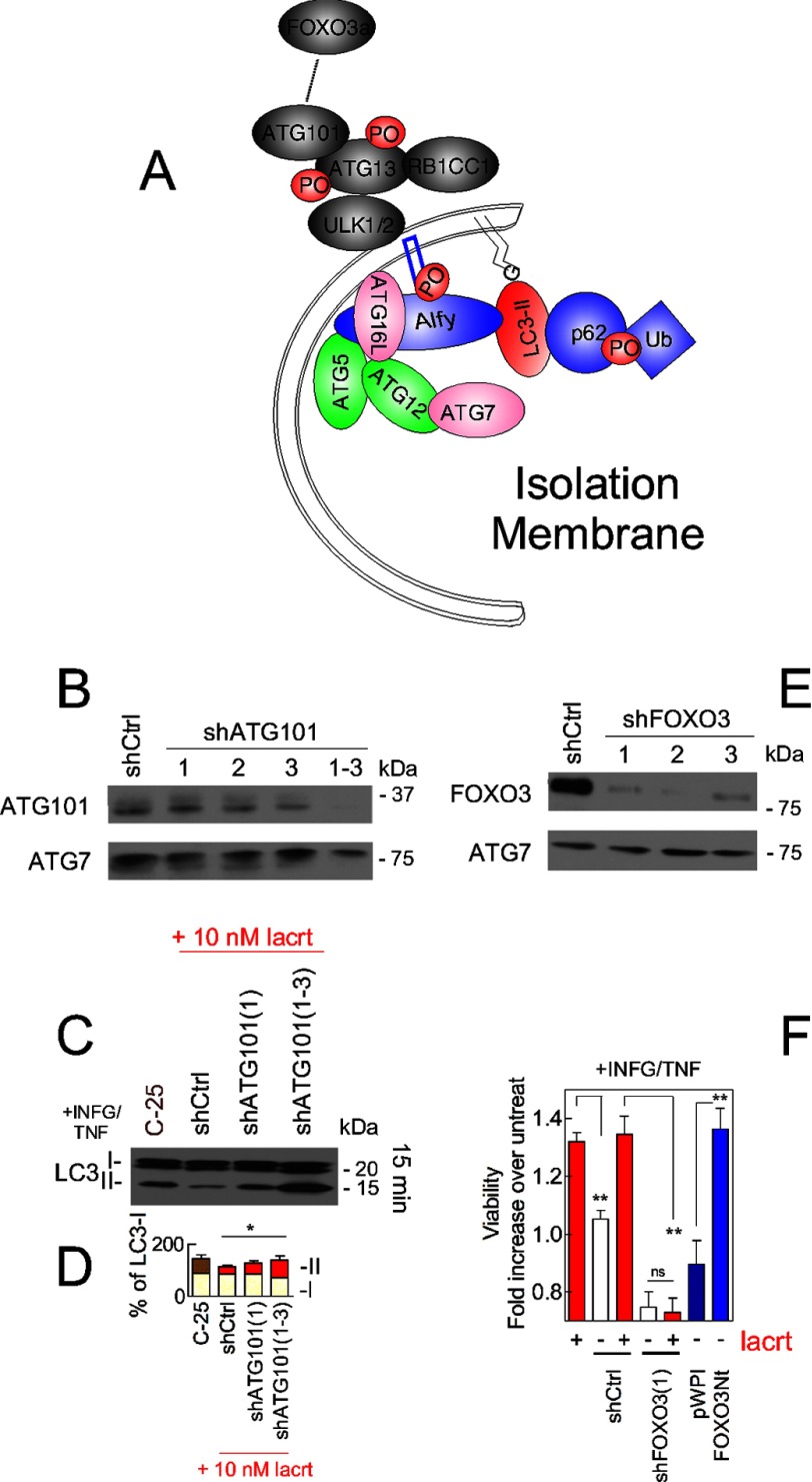
Homeostasis is essential for cell survival. However, homeostatic regulation of surface epithelia is poorly understood. The eye surface, lacking the cornified barrier of skin, provides an excellent model. Tears cover the surface of the eye and are deficient in dry eye, the most common eye disease affecting at least 5% of the world's population. Only a tiny fraction of the tear proteome appears to be affected, including lacritin, an epithelium-selective mitogen that promotes basal tearing when topically applied to rabbit eyes. Here we show that homeostasis of cultured corneal epithelia is entirely lacritin-dependent and elucidate the mechanism as a rapid autophagic flux to promptly restore cellular metabolism and mitochondrial fusion in keeping with the short residence time of lacritin on the eye. Accelerated flux appears to be derived from lacritin-stimulated acetylation of FOXO3 as a novel ligand for ATG101 and coupling of stress-acetylated FOXO1 with ATG7 (which remains uncoupled without lacritin) and be sufficient to selectively divert huntingtin mutant Htt103Q aggregates largely without affecting non-aggregated Htt25Q. This is in keeping with stress as a prerequisite for lacritin-stimulated autophagy. Lacritin targets the cell surface proteoglycan syndecan-1 via its C-terminal amino acids Leu(108)-Leu(109)-Phe(112) and is also available in saliva, plasma, and lung lavage. Thus, lacritin may promote epithelial homeostasis widely.

Keywords: ATG101; Autophagy; Cell Metabolism; Cornea; Dry Eye; FOXO3; Lacritin; Metabolomics; Proteoglycan; Stress.


Figures

FIGURE 1.
FIGURE 1.
Lacritin in normal human tears is essential for survival of stressed corneal epithelial cells. A, schematic diagram of normal tears on the eye surface as compared with skin with the protective cornified zone. B, schematic diagram of tears on the eye in dry eye. C, pool of basal human tears from 19 individuals. D, pool of dry eye human tears from 29 individuals. E, HCE-T cells were sensitized overnight with IFNG and then treated for 15 min with pooled normal tears in the presence of TNF. Cells were washed and fixed, and then FOXO3 was immunolocalized. The same approach with different tear or lacritin samples was used in F–J , M, and N. IFNG/TNF-stressed cells with lacritin-depleted tears (F), mock-depleted tears (G), pooled dry eye tears (H), dry eye tears spiked with 10 nm lacritin (I), and dry eye tears spiked with 10 nm C-25 (lacritin lacking 25 amino acids from the C terminus) (J). Bar, 10 μm. K, linear diagram of lacritin versus C-25. Yellow indicates demonstrated (15) and predicted α-helices. L, left panel, HCE-T cells were sensitized overnight with IFNG and then treated for 15 min with increasing amounts of lacritin (or FBS (middle panel)) in the presence of TNF. Viability was assessed by the MTT assay. 10 nm lacritin, but not other doses, significantly enhanced cell viability (ANOVA with Dunnett's post-test; **, p < 0.01) but is not significantly different from FBS-enhanced viability (p > 0.05). Right panel, HCE-T cells were similarly stressed with IFNG and TNF or not stressed and then left untreated or instead treated with 10 nm lacritin or C-25. Lacritin enhanced viability (ANOVA with Dunnett's post-test; **, p < 0.01). The y axis is not shown extending to zero in this and other viability assays because the assay detects subtle changes. M, lacritin (10 nm) on IFNG/TNF-stressed primary human corneal epithelial cells. Bar, 25 μm. N, C-25 (10 nm) on IFNG/TNF-stressed primary human corneal epithelial cells. Error bars represent S.E. cor, cornified; lacrt, lacritin; ep, epithelia; nml, normal.
FIGURE 2.
FIGURE 2.
Lacritin survival activity is defined by C-terminal hydrophobic residues Ile98, Phe104, Leu108, Leu109, and Phe112. A, pepwheel analysis of the C-terminal α-helix (amino acids 94–112) of lacritin. Red residues are hydrophobic. B, Predictor of Naturally Disordered Regions (PONDR) tracings of lacritin, lacritin mutants, and splice variant lacritin-c. 0 represents “order,” and 1 represents “disorder” with tracing below the horizontal line predicted to be ordered. The green rectangle approximates the syndecan-1 binding region (17) for cell targeting. C, linear diagrams comparing lacritin point (red ovals) and truncation mutants and splice variant with the novel I3 C terminus (blue). Numbering here and elsewhere excludes the signal peptide. D, HCE-T cells were sensitized overnight with IFNG and then treated for 15 min with different lacritin constructs (10 nm) in the presence of TNF. Viability was assessed by the MTT assay and is expressed as the -fold increase in viability, defined as the ratio of experimental viability to the control viability. Control viability was derived from cells treated with IFNG/TNF alone. Several mutants and the splice variant are not prosurvival (ANOVA with Dunnett's post-test; **, p < 0.01; *, p < 0.05). The viability of F104S, L108/L109/F112S, and lacritin I3-treated cells does not significantly differ from that of IFNG/TNF-stressed cells (ANOVA with Dunnett's post-test, ns, not significant, p > 0.05. E, HCE-T cells were sensitized overnight with IFNG in the presence of no or increasing amounts of xyloside (left panel). The next day, cells were treated for 15 min with 10 nm lacritin in the presence of TNF and xyloside. Viability was assessed by the MTT assay. Other cells were only treated overnight with no or increasing amounts of xyloside, and viability was assessed by the MTT assay (right panel). Xyloside significantly negates lacritin prosurvival activity. It also significantly reduces cell viability in the absence of IFNG/TNF but only slightly (94 ± 1.6 and 93 ± 1.9% versus 100% viability, respectively, for 80 and 90 μm xyloside). Xyloside inhibits heparan and chondroitin sulfate chain attachment and can inhibit hyaluronan synthesis. Left, diagrams of untreated (above) versus xyloside-treated (below) syndecan-1 with heparan and chondroitin sulfate side chains depicted in green and red, respectively. Both are attached to a core protein with a short cytoplasmic domain below the black line. Error bars represent S.E. lacrt, lacritin; SP, signal peptide; xyl, xyloside; aa, amino acids; untreat, untreated.
FIGURE 3.
FIGURE 3.
Stressed cells display enhanced autophagy that is further up-regulated by lacritin within 10 min. A, HCE-T cells were either not stressed or were stressed overnight with IFNG and then treated for 30 min without or with lacritin (10 nm) in the presence of TNF. Cell lysates were blotted for caspases (CASP) 9 and 3 or for LC3 and tubulin (loading control). LC3-I migrates more slowly than LC3-II. B, schematic diagram of autophagy with isolation membrane (left), autophagosome (center), autolysosome (right), and several autophagy mediators (LC3, ATG7, Alfy, and p62). HCE-T cells that stably express LC3 double tagged with EGFP and mCherry (22) were developed (LC3/RG cells) and used in G–M to monitor autophagic flux. Rectangles indicate the effect of pH on LC3 tags. C, HCE-T cells were sensitized overnight with IFNG and then treated for 6 h without or with TNF in the presence of no or increasing amounts of leupeptin (leu), vinblastine (vin), or wortmannin (wort). Lysates were blotted for LC3 or tubulin (loading control). D, autophagic flux is chronically enhanced by TNF stress as revealed by accumulation of LC3-II with leupeptin and vinblastine. Each value plotted is the mean integrated optical density of the films (n = 3) normalized to the tubulin loading control. E, HCE-T cells were sensitized overnight with IFNG and then treated for 30 min with lacritin (10 nm) in the presence of TNF without or with 0.1 μm leupeptin. Lysates were blotted for LC3 or tubulin (loading control). F, quantitation of replicate experiments from E (paired t test for LC3-II; *, p < 0.05) plotted as indicated in D. LC3/RG cells were sensitized overnight with IFNG and then treated for different times with 10 nm lacritin (G) or C-25 in the presence of TNF without (H) or with 0.05 μm vinblastine (vinblast) (I). Puncta per field in microscopic images were quantitated (ANOVA with Dunnett's post-test; **, p < 0.01; *, p < 0.05; ns, not significant). Representative microscopic images are shown in J–M. J, C-25 for 60 min. K, lacritin for 60 min. C-25 (L) and lacritin (M) treatment for 60 min in the presence of vinblastine (Vin; 0.05 μm) is shown. Bar, 10 μm. Error bars represent S.E.
FIGURE 4.
FIGURE 4.
Lacritin-stimulated autophagy is calcineurin- but not mTOR-dependent and necessary for cell survival. A, HCE-T cells were either not stressed or alternatively were stressed overnight with IFNG and then treated for 15 or 120 min with 10 nm lacritin or C-25 in the absence or presence of TNF. Cell lysates were blotted for phospho-S6K or S6K (loading control). Phospho-S6K is an indicator of mTOR activity. B, quantitation of replicate experiments from A (paired t test; ns, not significant, p > 0.05). Each value plotted is the mean integrated optical density of the films (n = 3) normalized to the S6K loading control. C, HCE-T cells were stressed overnight with IFNG and then treated for 15 min with 10 nm lacritin in the presence of TNF without or with PI103, rapamycin (Rap), cyclosporin A (CsA), or rapamycin plus cyclosporin A. Viability was assessed by the MTT assay and is expressed as the -fold increase in viability, defined as the ratio of experimental viability to the control viability. Control viability was derived from cells treated with IFNG/TNF alone (ANOVA with Dunnett's post-test; **, p < 0.01). Viability with 0.1 μm cyclosporin A or 0.1 μm cyclosporin A and rapamycin treatment plus IFNG/TNF and lacritin does not significantly differ from viability of cells treated with IFNG/TNF alone (ANOVA with Dunnett's post-test; ns, not significant, p > 0.05). D, HCE-T cells stably expressing empty vector (shCtrl) or three different ATG7 shRNAs were developed. Each was stressed overnight with IFNG and then treated for 15 min with 10 nm lacritin in the presence of TNF. Viability was assessed by the MTT assay and is expressed as the -fold increase in viability as defined above (ANOVA with Dunnett's post-test; **, p < 0.01). Viability from shATG7(3) cells with IFNG/TNF and lacritin differs significantly from shCtrl cells treated with IFNG/TNF alone (t test; *, p = 0.03); thus, shATG7(1) was used in subsequent experiments. E, Western blot for ATG7 in shCtrl and shATG7 knockdown cell lines using FOXO3 as a loading control. F, shCtrl or shATG7(1) HCE-T cells were sensitized overnight with IFNG and then treated for 15 min with lacritin (10 nm) in the presence of TNF. Cells were washed and fixed, and then FOXO3 was immunolocalized. G, HCE-T cells were sensitized overnight with IFNG and then treated for different times with 10 nm lacritin (top pair) or C-25 (bottom pair) in the presence of TNF. Lysates were blotted for LC3 or tubulin (loading control). H, quantitation of G replicates plotted as indicated in B normalized to the tubulin loading control. I, HCE-T cells were sensitized overnight with IFNG and then treated for 15 min with different doses of lacritin (top pair) or C-25 (bottom pair) in the presence of TNF. Lysates were blotted for LC3 or tubulin. J, quantitation of I replicates plotted as indicated in H. H and J, two-way ANOVA (LC3-II and LC3-I); *, p < 0.05. Error bars represent S.E. Inhib, inhibitor; lacrt, lacritin; pS6K, phospho-S6K.
FIGURE 5.
FIGURE 5.
Lacritin-stimulated autophagy captures Htt103Q cellular aggregates. A, Htt103Q-mCFP or Htt25Q-mCFP constructs transduced in HCE-T cells stably expressing LC3 double tagged with EGFP and mCherry (22) (LC3/RG cells). B, quantitation of colocalized mCFP-Htt and mCherry LC3 at different times after treatment with 10 nm lacritin or C-25 in the absence of IFNG and TNF (t test; **, p < 0.01). C, number of LC3 mCherry- or LC3 mCherry-EGFP-labeled puncta in cells expressing Htt103Q or Htt25Q at different times after treatment with 10 nm lacritin or C-25 (no IFNG/TNF) (t test; *, p < 0.05). D, number of Htt103Q or Htt25Q puncta greater or less than 1 μm at different times after treatment with 10 nm lacritin or C-25 (no IFNG/TNF) (t test; *, p < 0.05; **, p < 0.01). Representative microscopic images are shown in E–H. E and F, LC3/RG cells expressing Htt103Q-mCFP that were treated with lacritin (E) or C-25 (F) for different times. Cells were washed and fixed, and all tags were localized. IFNG/TNF was omitted. G and H, LC3/RG cells expressing Htt25Q-mCFP that were treated with lacritin (G) or C-25 (H) for different times. IFNG/TNF was omitted. Bar, 10 μm. Error bars represent S.E. lacrt, lacritin.
FIGURE 6.
FIGURE 6.
Lacritin-stimulated autophagic capture with p62 and Alfy. A, HCE-T cells were stressed overnight with IFNG and then treated for different times with 10 nm lacritin or for 30 min with 10 nm C-25 or EGF (1 μg/ml) in the presence of TNF. LC3 was immunoprecipitated (IP) from cell lysates. Immunoprecipitated material was blotted for Alfy, p62, or ubiquitin. B, same as A in which cells were treated with 10 nm C-25 for different times. C–E, quantitation of A and B with replicates (ANOVA with Dunnett's post-test; *, p < 0.05; **, p < 0.01). Each value plotted is the mean integrated optical density of the films (n = 3). lacrt, lacritin.
FIGURE 7.
FIGURE 7.
Lacritin restores metabolism. A, HCE-T cells were stressed overnight with IFNG and then treated for different times with 10 nm lacritin or C-25 in the presence of TNF. The oxygen consumption rate was monitored in the absence or presence of oligomycin (500 nm), FCCP (375 nm), or antimycin A (750 nm) and rotenone (1 μm) together. B, features of the oxygen consumption rate as revealed by use of oligomycin, FCCP, antimycin A, and rotenone (t test; **, p < 0.01). C, HCE-T or HCE-T shATG7(1) cells were stressed overnight with IFNG and then stained for 30 min with MitoTracker Red FM. After addition of lacritin or C-25 (10 nm) in the presence of TNF, live mitochondria were monitored at 20-s intervals for 30 min, and fission frequency was estimated from ComponentCount of Mytoe. D, schematic diagram of mitochondrial fusion or fission. E, HCE-T cells were stressed overnight with IFNG and then treated for 10 min in replicates of six with 10 nm lacritin or C-25 or left untreated in the presence of TNF. Metabolites in cell lysates were identified by mass spectrometry. Values were analyzed by the t test and non-parametric Wilcoxon test of the log-transformed normalized data (*, p < 0.05) and further analyzed by hierarchical clustering. Error bars represent S.E. lacrt, lacritin; antimyc, antimycin A; rot, rotenone; res, respiration; res cap, respiratory capacity; untreat, untreated.
FIGURE 8.
FIGURE 8.
Lacritin stimulated FOXO1-ATG7 and FOXO3-ATG101 coupling. A, HCE-T cells were either not stressed or stressed overnight with IFNG and treated for different times with TNF. FOXO1 was immunoprecipitated (IP) from cell lysates, and the immunoprecipitated material was blotted for acetyl-lysine, ATG7, or FOXO1 (loading control). B, quantitation from replicate blots (ANOVA with Dunnett's post-test; *, p < 0.05; **, p < 0.01). Each value plotted is the mean integrated optical density of the films (n = 3) normalized to the FOXO1 loading control. C, same as A in which stressed cells were also treated with 10 nm lacritin or C-25. D, quantitation from replicate blots (ANOVA with Dunnett's post-test; *, p < 0.05; **, p < 0.01) plotted as indicated in B. E, replicate blot of the 15-min time point from C without or with lacritin. F, quantitation of replicate blots from E plotted as indicated in B (t test; **, p < 0.01). G, HCE-T cells were either not stressed or stressed overnight with IFNG and treated for different times with TNF. FOXO3 was immunoprecipitated from cell lysates, and the immunoprecipitate was blotted for acetyl-lysine, ATG7, or FOXO3 (loading control). H, quantitation from replicate blots (ANOVA; **, p < 0.01) plotted as indicated in B but normalized to the FOXO3 loading control. I, HCE-T cells were either not stressed or stressed overnight with IFNG and treated for different times with 10 nm lacritin in the presence of TNF. FOXO3 was immunoprecipitated from cell lysates. Immunoprecipitated material was blotted for ATG101 or FOXO3 (loading control). J, replicate blot of the 15-min time point from I with 10 nm lacritin, C-25, or lysate alone. K, quantitation from replicate blots (ANOVA with Dunnett's post-test; *, p < 0.05; **, p < 0.01) plotted as indicated in H. Error bars represent S.E. lacrt, lacritin; ac-lys, acetyl-lysine.
FIGURE 9.
FIGURE 9.
FOXO3 and ATG101 are essential for lacritin stimulated autophagy. A, schematic diagram of the isolation membrane illustrating several associated autophagic mediators. B, ATG101 immunoblot of stably transduced shCtrl, shATG101(1), shATG101(2), shATG101(3), or shATG101(1–3) HCE-T cells. ATG7 was used as a loading control. C, HCE-T and stably transduced HCE-T shCtrl, shATG101(1), and shATG101(1–3) cells were stressed overnight with IFNG and then treated for 15 min with 10 nm lacritin or C-25 in the presence of TNF. Lysates were blotted for LC3. D, quantitation of replicates from B (ANOVA for LC3-II; *, p < 0.05). Each value plotted is the mean integrated optical density of the films (n = 3) normalized to the tubulin loading control. E, FOXO3 immunoblot of stably transduced shCtrl, shFOXO3(1), shFOXO3(2), and shFOXO3(3) cells. ATG7 was used as a loading control. F, HCE-T, shCtrl, shFOXO3(1), pWPI, and FOXO3Nt cells were stressed overnight with IFNG and incubated for 15 min with TNF in the absence or presence of 10 nm lacritin. For comparison, HCE-T cells were transduced with pWPI vector or with FOXO3Nt (in pWPI). Cells were stressed overnight with IFNG and incubated for 15 min with TNF without lacritin. Viability was assessed by the MTT assay and is expressed as the -fold increase in viability, defined as the ratio of experimental viability to the control viability. Control viability was derived from cells treated with IFNG/TNF alone (t test; **, p < 0.01; ns, not significant, p > 0.05). Error bars represent S.E. lacrt, lacritin; Ub, ubiquitin; untreat, untreated.

Similar articles

Cited by

  • The Impact of Aging on Ocular Diseases: Unveiling Complex Interactions.
    You L, Lin Y, Zheng Y, Han Z, Zeng L, Chen H. You L, et al. Aging Dis. 2024 Sep 23;16(5):2803-2830. doi: 10.14336/AD.2024.0850. Aging Dis. 2024. PMID: 39500360 Free PMC article. Review.
  • Guidelines for the use and interpretation of assays for monitoring autophagy (4th edition)1.
    Klionsky DJ, Abdel-Aziz AK, Abdelfatah S, Abdellatif M, Abdoli A, Abel S, Abeliovich H, Abildgaard MH, Abudu YP, Acevedo-Arozena A, Adamopoulos IE, Adeli K, Adolph TE, Adornetto A, Aflaki E, Agam G, Agarwal A, Aggarwal BB, Agnello M, Agostinis P, Agrewala JN, Agrotis A, Aguilar PV, Ahmad ST, Ahmed ZM, Ahumada-Castro U, Aits S, Aizawa S, Akkoc Y, Akoumianaki T, Akpinar HA, Al-Abd AM, Al-Akra L, Al-Gharaibeh A, Alaoui-Jamali MA, Alberti S, Alcocer-Gómez E, Alessandri C, Ali M, Alim Al-Bari MA, Aliwaini S, Alizadeh J, Almacellas E, Almasan A, Alonso A, Alonso GD, Altan-Bonnet N, Altieri DC, Álvarez ÉMC, Alves S, Alves da Costa C, Alzaharna MM, Amadio M, Amantini C, Amaral C, Ambrosio S, Amer AO, Ammanathan V, An Z, Andersen SU, Andrabi SA, Andrade-Silva M, Andres AM, Angelini S, Ann D, Anozie UC, Ansari MY, Antas P, Antebi A, Antón Z, Anwar T, Apetoh L, Apostolova N, Araki T, Araki Y, Arasaki K, Araújo WL, Araya J, Arden C, Arévalo MA, Arguelles S, Arias E, Arikkath J, Arimoto H, Ariosa AR, Armstrong-James D, Arnauné-Pelloquin L, Aroca A, Arroyo DS, Arsov I, Artero R, Asaro DML, Aschner M, Ashrafizadeh M, Ashur-Fabian O, Atanasov AG, Au AK, Auberger P, Auner HW, Aurelian L, Autelli R… See abstract for full author list ➔ Klionsky DJ, et al. Autophagy. 2021 Jan;17(1):1-382. doi: 10.1080/15548627.2020.1797280. Epub 2021 Feb 8. Autophagy. 2021. PMID: 33634751 Free PMC article.
  • Topical administration of lacritin is a novel therapy for aqueous-deficient dry eye disease.
    Vijmasi T, Chen FY, Balasubbu S, Gallup M, McKown RL, Laurie GW, McNamara NA. Vijmasi T, et al. Invest Ophthalmol Vis Sci. 2014 Jul 17;55(8):5401-9. doi: 10.1167/iovs.14-13924. Invest Ophthalmol Vis Sci. 2014. PMID: 25034600 Free PMC article.
  • A cleavage-potentiated fragment of tear lacritin is bactericidal.
    McKown RL, Coleman Frazier EV, Zadrozny KK, Deleault AM, Raab RW, Ryan DS, Sia RK, Lee JK, Laurie GW. McKown RL, et al. J Biol Chem. 2014 Aug 8;289(32):22172-82. doi: 10.1074/jbc.M114.570143. Epub 2014 Jun 18. J Biol Chem. 2014. PMID: 24942736 Free PMC article.
  • Targeting Iron - Respiratory Reciprocity Promotes Bacterial Death.
    Sharifian Gh M, Norouzi F, Sorci M, Zaid TS, Pier GB, Achimovich A, Ongwae GM, Liang B, Ryan M, Lemke M, Belfort G, Gadjeva M, Gahlmann A, Pires MM, Venter H, Harris TE, Laurie GW. Sharifian Gh M, et al. bioRxiv [Preprint]. 2024 Mar 1:2024.03.01.582947. doi: 10.1101/2024.03.01.582947. bioRxiv. 2024. Update in: J Biol Chem. 2025 May;301(5):108455. doi: 10.1016/j.jbc.2025.108455. PMID: 38464199 Free PMC article. Updated. Preprint.

References

    1. Morimoto R. I. (2008) Proteotoxic stress and inducible chaperone networks in neurodegenerative disease and aging. Genes Dev. 22, 1427–1438 - PMC - PubMed
    1. Levine B., Mizushima N., Virgin H. W. (2011) Autophagy in immunity and inflammation. Nature 469, 323–335 - PMC - PubMed
    1. Hotamisligil G. S. (2006) Inflammation and metabolic disorders. Nature 444, 860–867 - PubMed
    1. Lamark T., Johansen T. (2010) Autophagy: links with the proteasome. Curr. Opin. Cell Biol. 22, 192–198 - PubMed
    1. Zhao Y., Yang J., Liao W., Liu X., Zhang H., Wang S., Wang D., Feng J., Yu L., Zhu W. G. (2010) Cytosolic FOXO1 is essential for the induction of autophagy and tumour suppressor activity. Nat. Cell Biol. 12, 665–675 - PubMed

Publication types

MeSH terms