

Cerebral TOF angiography at 7T: Impact of B1 (+) shimming with a 16-channel transceiver array
- PMID: 23640915
- PMCID: PMC4097948
- DOI: 10.1002/mrm.24749
Cerebral TOF angiography at 7T: Impact of B1 (+) shimming with a 16-channel transceiver array
Abstract
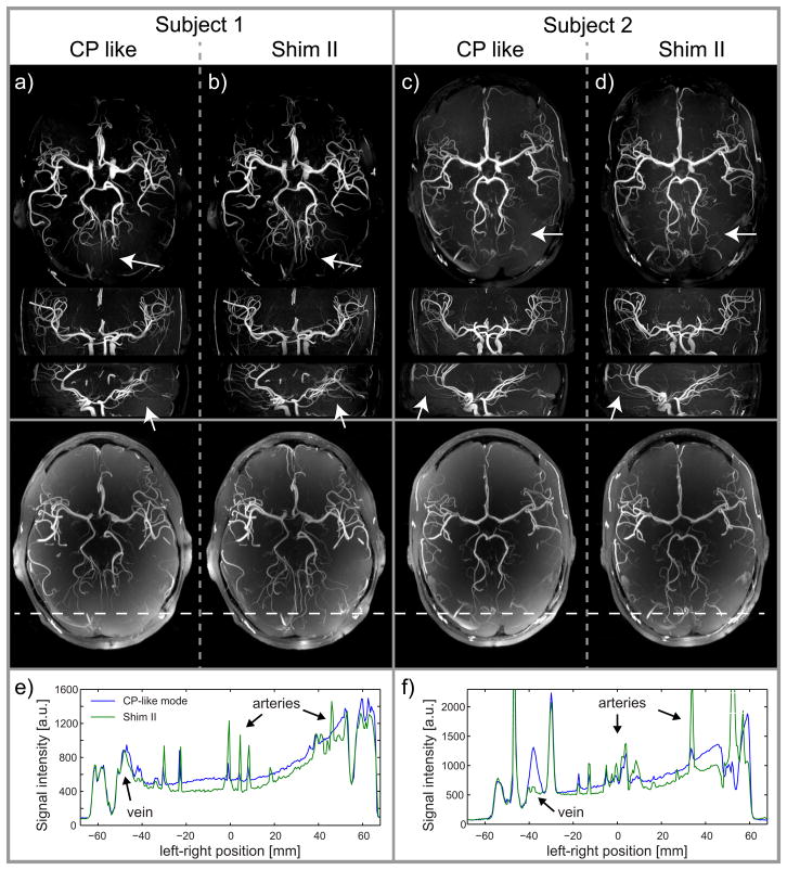
Purpose: Time-of-flight (TOF) MR imaging is clinically among the most common cerebral noncontrast enhanced MR angiography techniques allowing for high spatial resolution. As shown by several groups TOF contrast significantly improves at ultrahigh field of B0 = 7T, however, spatially varying transmit B1 (B1 (+)) fields at 7T reduce TOF contrast uniformity, typically resulting in suboptimal contrast and reduced vessel conspicuity in the brain periphery.
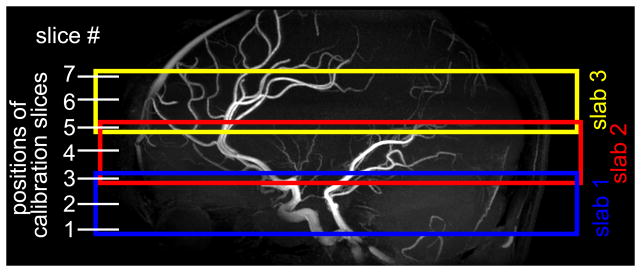
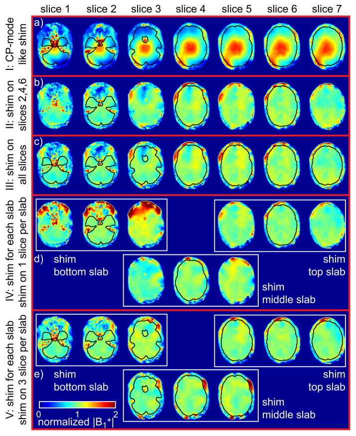
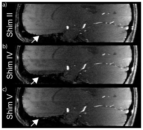
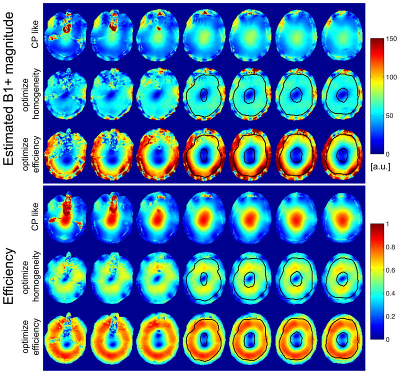
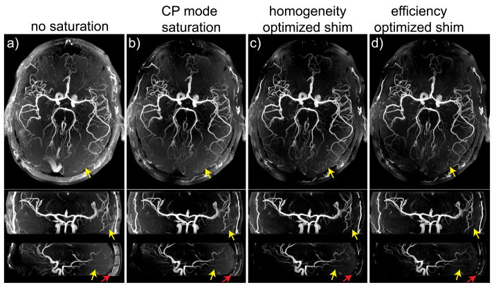
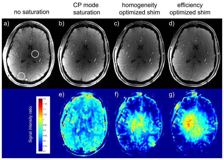
Methods: Using a 16-channel B1 (+) shimming system, we compare different dynamically applied B1 (+) phase shimming approaches on the radiofrequency excitation to improve contrast homogeneity for a (0.5 mm)(3) resolution multislab TOF acquisition. In addition, B1 (+) shimming applied on the venous saturation pulse was investigated to improve venous suppression, subcutaneous fat signal reduction and enhanced background suppression originating from MT effect.
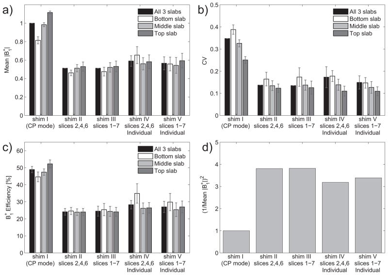
Results: B1 (+) excitation homogeneity was improved by a factor 2.2-2.6 on average depending on the shimming approach, compared to a standard CP-like phase setting, leading to improved vessel conspicuity particularly in the periphery. Stronger saturation, higher fat suppression and improved background suppression were observed when dynamically applying B1 (+) shimming on the venous saturation pulse.
Conclusion: B1+ shimming can significantly improve high resolution TOF vascular investigations at ultrahigh field, holding strong promise for non contrast-enhanced clinical applications.
Copyright © 2013 Wiley Periodicals, Inc.
Figures
Similar articles
-
Seven-tesla time-of-flight angiography using a 16-channel parallel transmit system with power-constrained 3-dimensional spoke radiofrequency pulse design.Invest Radiol. 2014 May;49(5):314-25. doi: 10.1097/RLI.0000000000000033. Invest Radiol. 2014. PMID: 24598439 Free PMC article.
-
Contrast enhancement in TOF cerebral angiography at 7 T using saturation and MT pulses under SAR constraints: impact of VERSE and sparse pulses.Magn Reson Med. 2012 Jul;68(1):188-97. doi: 10.1002/mrm.23226. Epub 2011 Dec 2. Magn Reson Med. 2012. PMID: 22139829 Free PMC article.
-
Cardiac imaging at 7 Tesla: Single- and two-spoke radiofrequency pulse design with 16-channel parallel excitation.Magn Reson Med. 2013 Nov;70(5):1210-9. doi: 10.1002/mrm.24935. Epub 2013 Sep 10. Magn Reson Med. 2013. PMID: 24038314 Free PMC article.
-
[Basic principles of MR angiography. An introduction].Radiologe. 1994 Aug;34(8):416-22. Radiologe. 1994. PMID: 7972718 Review. German.
-
Cerebrovascular MRI: a review of state-of-the-art approaches, methods and techniques.NMR Biomed. 2015 Jul;28(7):767-91. doi: 10.1002/nbm.3322. Epub 2015 May 26. NMR Biomed. 2015. PMID: 26010775 Review.
Cited by
-
Towards high-resolution 4D flow MRI in the human aorta using kt-GRAPPA and B1+ shimming at 7T.J Magn Reson Imaging. 2016 Aug;44(2):486-99. doi: 10.1002/jmri.25164. Epub 2016 Feb 3. J Magn Reson Imaging. 2016. PMID: 26841070 Free PMC article.
-
A new RF transmit coil for foot and ankle imaging at 7T MRI.Magn Reson Imaging. 2018 Jan;45:1-6. doi: 10.1016/j.mri.2017.09.005. Epub 2017 Sep 8. Magn Reson Imaging. 2018. PMID: 28893660 Free PMC article.
-
Prospective motion correction enables highest resolution time-of-flight angiography at 7T.Magn Reson Med. 2018 Jul;80(1):248-258. doi: 10.1002/mrm.27033. Epub 2017 Dec 11. Magn Reson Med. 2018. PMID: 29230871 Free PMC article.
-
Magnetic resonance imaging at ultrahigh fields.IEEE Trans Biomed Eng. 2014 May;61(5):1364-79. doi: 10.1109/TBME.2014.2313619. Epub 2014 Mar 25. IEEE Trans Biomed Eng. 2014. PMID: 24686229 Free PMC article. Review.
-
Comparison of RF body coils for MRI at 3 T: a simulation study using parallel transmission on various anatomical targets.NMR Biomed. 2015 Oct;28(10):1332-44. doi: 10.1002/nbm.3378. Epub 2015 Aug 30. NMR Biomed. 2015. PMID: 26332290 Free PMC article.
References
-
- Heverhagen JT, Bourekas E, Sammet S, Knopp MV, Schmalbrock P. Time-of-flight magnetic resonance angiography at 7 Tesla. Invest Radiol. 2008;43(8):568–573. - PubMed
-
- Kang CK, Park CW, Han JY, Kim SH, Park CA, Kim KN, Hong SM, Kim YB, Lee KH, Cho ZH. Imaging and analysis of lenticulostriate arteries using 7. 0-Tesla magnetic resonance angiography. Magn Reson Med. 2009;61(1):136–144. - PubMed
-
- Zwanenburg JJ, Hendrikse J, Takahara T, Visser F, Luijten PR. MR angiography of the cerebral perforating arteries with magnetization prepared anatomical reference at 7 T: comparison with time-of-flight. J Magn Reson Imaging. 2008;28(6):1519–1526. - PubMed
-
- Rooney WD, Johnson G, Li X, Cohen ER, Kim SG, Ugurbil K, Springer CS., Jr Magnetic field and tissue dependencies of human brain longitudinal 1H2O relaxation in vivo. Magn Reson Med. 2007;57(2):308–318. - PubMed
Publication types
MeSH terms
Grants and funding
LinkOut - more resources
Full Text Sources
Other Literature Sources
Research Materials
Miscellaneous
