. 2013 Jul;61(7):487-99.
doi: 10.1369/0022155413490947. Epub 2013 May 2.

Diaphragm muscle remodeling in a rat model of chronic intermittent hypoxia

Affiliations

Diaphragm muscle remodeling in a rat model of chronic intermittent hypoxia

Christine M Shortt et al. J Histochem Cytochem. 2013 Jul.

Abstract

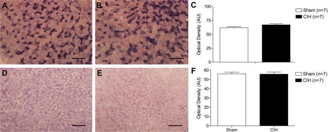
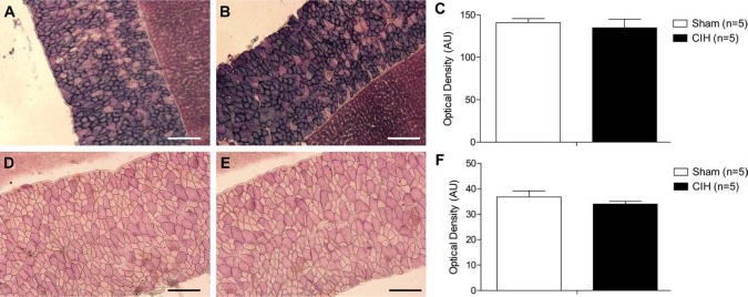
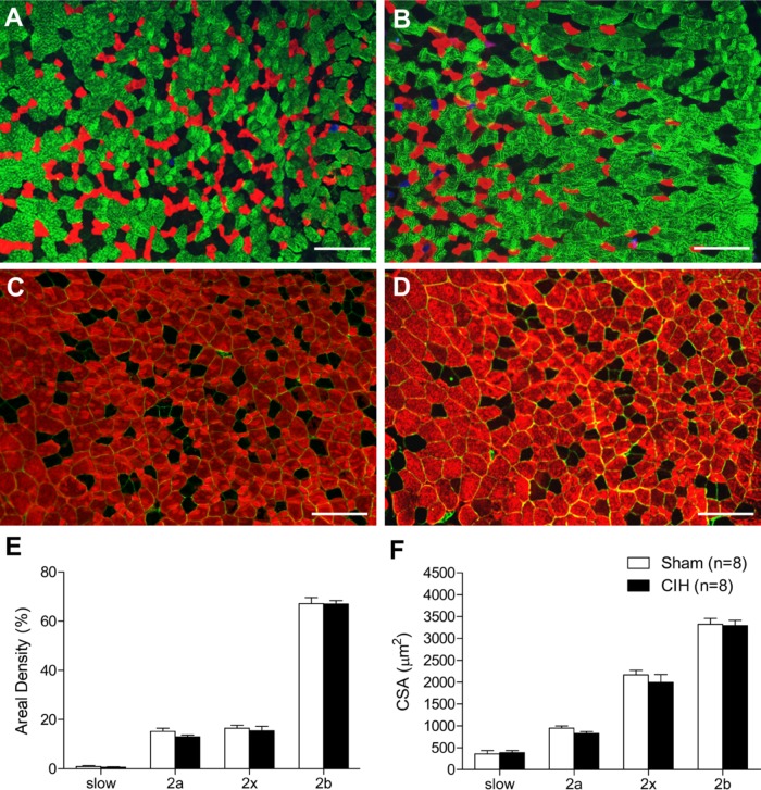
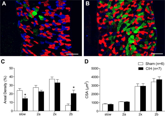
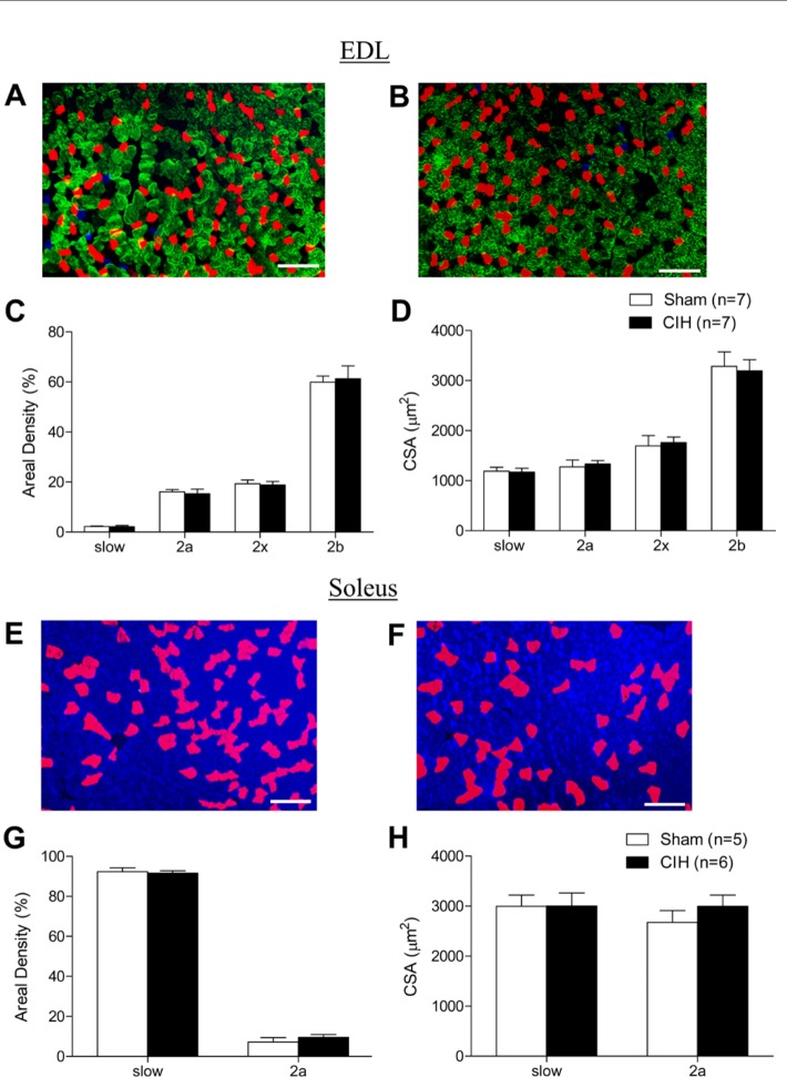
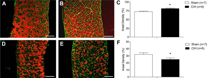
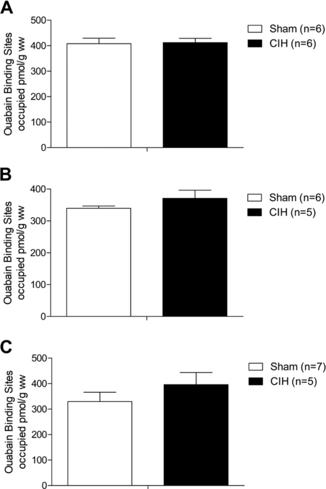
Respiratory muscle remodeling occurs in human sleep apnea--a common respiratory disorder characterized by chronic intermittent hypoxia (CIH) due to recurrent apnea during sleep. We sought to determine if CIH causes remodeling in rat sternohyoid (upper airway dilator) and diaphragm muscles. Adult male Wistar rats were exposed to CIH (n=8), consisting of 90 sec of hypoxia (5% at the nadir; SaO₂ ~80%)/90 sec of normoxia, 8 hr per day, for 7 consecutive days. Sham animals (n=8) were exposed to alternating air/air cycles in parallel. The effect of CIH on myosin heavy-chain (MHC) isoform (1, 2a, 2x, 2b) distribution, sarcoplasmic reticulum calcium ATPase (SERCA) isoform distribution, succinate dehydrogenase activity, glycerol phosphate dehydrogenase activity, and Na⁺/K⁺ ATPase pump content was determined. Sternohyoid muscle structure was unaffected by CIH treatment. CIH did not alter oxidative/glycolytic capacity or the Na⁺/K⁺-ATPase pump content of the diaphragm. CIH significantly increased the areal density of MHC 2b fibers in the rat diaphragm, and this was associated with a shift in SERCA proteins from SERCA2 to SERCA1. We conclude that CIH causes a slow-to-fast fiber transition in the rat diaphragm after just 7 days of treatment. Respiratory muscle functional remodeling may drive aberrant functional plasticity such as decreased muscle endurance, which is a feature of human sleep apnea.

Keywords: chronic intermittent hypoxia; diaphragm dysfunction; myosin heavy chain isoform distribution; obstructive sleep apnea; respiratory muscles.


Conflict of interest statement

Declaration of Conflicting Interests: The author(s) declared no potential conflicts of interest with respect to the research, authorship, and/or publication of this article.

Figures

Figure 1.
Figure 1.
Sternohyoid enzymatic activity. Representative images of sternohyoid muscle stained for succinate dehydrogenase (SDH)—an index of enzymatic oxidative capacity in sternohyoid muscle from a sham (A) and chronic intermittent hypoxia (CIH)-treated (B) animal. (C) Group data (mean ± SEM) showing that CIH had no effect on sternohyoid SDH activity. Representative images of glycerol phosphate dehydrogenase (GPDH) staining (a marker of glycolytic enzymatic activity) in sternohyoid muscle from a sham (D) and CIH-treated (E) animal. (F) Group data (mean ± SEM) showing that CIH had no effect on sternohyoid GPDH activity. Bars = 200 µm.
Figure 2.
Figure 2.
Diaphragm enzymatic activity. Representative images of diaphragm muscle (and an adjacent piece of liver used to assist transverse cutting of the tissue) stained for succinate dehydrogenase (SDH)—an index of enzymatic oxidative capacity in a sham (A) and chronic intermittent hypoxia (CIH)–treated (B) animal. (C) Group data (mean ± SEM) showing that CIH had no effect on diaphragm SDH activity. Representative images of glycerol phosphate dehydrogenase (GPDH) staining (a marker of glycolytic enzymatic activity) in diaphragm muscle (and an adjacent piece of liver used to assist transverse cutting of the tissue) from a sham (D) and CIH-treated (E) animal. (F) Group data (mean ± SEM) showing that CIH had no effect on diaphragm GPDH activity. Bars = 200 µm.
Figure 3.
Figure 3.
Sternohyoid myosin heavy-chain (MHC) areal density and fiber cross-sectional area (CSA). Representative images of sternohyoid muscle in a sham (A) and chronic intermittent hypoxia (CIH)–treated (B) animal triple-labeled with monoclonal antibodies showing MHC 1 (blue), MHC 2a (red), and MHC 2b (green); MHC 2x fibers are untagged. Representative images of sternohyoid muscle in a sham (C) and CIH-treated (D) animal double-labeled with monoclonal antibodies tagging the basement membrane protein laminin (green) and all MHC isoforms (red) except for MHC 2x (untagged). (E) Group data (mean ± SEM) showing that CIH had no significant effect on the areal density of sternohyoid muscle MHC 1 (slow), MHC 2a, MHC 2x, or MHC 2b fibers. (F) Group data (mean ± SEM) showing that CIH had no significant effect on sternohyoid fiber CSAs. Bars = 200 µm.
Figure 4.
Figure 4.
Diaphragm myosin heavy-chain (MHC) areal density and fiber cross-sectional area (CSA). Representative images of diaphragm muscle from a sham (A) and chronic intermittent hypoxia (CIH)–treated animal (B) labeled with monoclonal antibodies showing MHC 1 (blue), MHC 2a (red), and MHC 2b (green); MHC 2x fibers are untagged. Note the decrease in MHC 1 (blue) and large increase in MHC 2b (green) fibers in the CIH-treated animal (B) compared with the sham animal (A). (C) Group data (mean ± SEM) showing that CIH significantly decreased MHC 1 (slow) areal density (*p=0.006, Student’s unpaired t-test) and increased MHC 2b areal density (*p=0.008) in the diaphragm. MHC 2a and MHC 2x areal densities were not statistically different in sham and CIH-treated animals (C). (D) Group data (mean ± SEM) showing that CIH had no significant effect on diaphragm fiber CSAs. Bars = 200 µm.
Figure 5.
Figure 5.
Limb myosin heavy-chain (MHC) areal density and fiber cross-sectional area (CSA). Representative images of extensor digitorum longus (EDL) muscle from a sham (A) and chronic intermittent hypoxia (CIH)–treated (B) animal labeled with monoclonal antibodies showing MHC 1 (blue), MHC 2a (red), and MHC 2b (green); MHC 2x fibers are untagged. (C) Group data (mean ± SEM) showing that CIH had no significant effect on the areal density of MHC 1 (slow), MHC 2a, MHC 2x, or MHC 2b EDL muscle fibers. (D) Group data (mean ± SEM) showing that CIH had no significant effect on EDL fiber CSAs. Representative images of soleus muscle from a sham (E) and CIH-treated (F) animal labeled with monoclonal antibodies showing MHC 1 (blue) and MHC 2a (red). (G) Group data (mean ± SEM) showing that CIH had no significant effect on the areal density of MHC 1 (slow) or MHC 2a soleus muscle fibers. (H) Group data (mean ± SEM) showing that CIH had no significant effect on soleus fiber CSAs. Bars = 200 µm.
Figure 6.
Figure 6.
Diaphragm sarcoplasmic reticulum calcium ATPase (SERCA) isoform distribution. Representative images of diaphragm muscle from a sham (A) and chronic intermittent hypoxia (CIH)–treated (B) animal positively labeled using antibodies for SERCA1 isoform (red) and laminin (green). Note the increase in SERCA1 isoform distribution in the CIH-treated animal (B) compared with the sham animal (A). (C) Group data (mean ± SEM) show that CIH significantly increased SERCA1 isoform distribution compared with sham (*p=0.004, Student’s unpaired t-test). Representative images of diaphragm muscle from a sham (D) and CIH-treated (E) animal positively labeled using antibodies for SERCA2 isoform (red) and laminin (green). Note the decrease in SERCA2 isoform distribution in the CIH-treated animal (E) compared with the sham animal (D). (F) Group data (mean ± SEM) shows that SERCA2 isoform distribution was significantly decreased in CIH-treated animals compared with sham animals (*p=0.05, Student’s unpaired t-test). Bars = 200 µm.
Figure 7.
Figure 7.
Na+/K+-ATPase pump content in skeletal muscle. Group data (mean ± SEM) show that chronic intermittent hypoxia (CIH) had no significant effect on diaphragm (A), soleus (B), or extensor digitorum longus (C) Na+/K+-ATPase pump content.

Similar articles

Cited by

References

    1. Allen DG, Lamb GD, Westerblad H. 2008. Skeletal muscle fatigue: cellular mechanisms. Physiol Rev. 88:287–332 - PubMed
    1. Aubier M, Viires N. 1998. Calcium ATPase and respiratory muscle function. Eur Respir J. 11:758–766 - PubMed
    1. Aughey RJ, Gore CJ, Hahn AG, Garnham AP, Clark SA, Petersen AC, Roberts AD, McKenna MJ. 2005. Chronic intermittent hypoxia and incremental cycling exercise independently depress muscle in vitro maximal Na+-K+-ATPase activity in well-trained athletes. J Appl Physiol. 98:186–192 - PubMed
    1. Baldwin KM, Haddad F. 2002. Skeletal muscle plasticity: cellular and molecular responses to altered physical activity paradigms. Am J Phys Med Rehabil. 81:S40–S51 - PubMed
    1. Bradford A. 2004. Effects of chronic intermittent asphyxia on haematocrit, pulmonary arterial pressure and skeletal muscle structure in rats. Exp Physiol. 89:44–52 - PubMed

Publication types

Substances