

Fatal neurological respiratory insufficiency is common among viral encephalitides
- PMID: 23641019
- PMCID: PMC3719899
- DOI: 10.1093/infdis/jit186
Fatal neurological respiratory insufficiency is common among viral encephalitides
Abstract
Background: Neurological respiratory insufficiency strongly correlates with mortality among rodents infected with West Nile virus (WNV), which suggests that this is a primary mechanism of death in rodents and possibly fatal West Nile neurological disease in human patients.
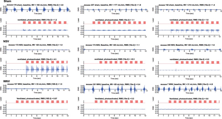
Methods: To explore the possibility that neurological respiratory insufficiency is a broad mechanism of death in cases of viral encephalitis, plethysmography was evaluated in mice infected with 3 flaviviruses and 2 alphaviruses. Pathology was investigated by challenging the diaphragm, using electromyography with hypercapnia and optogenetic photoactivation.
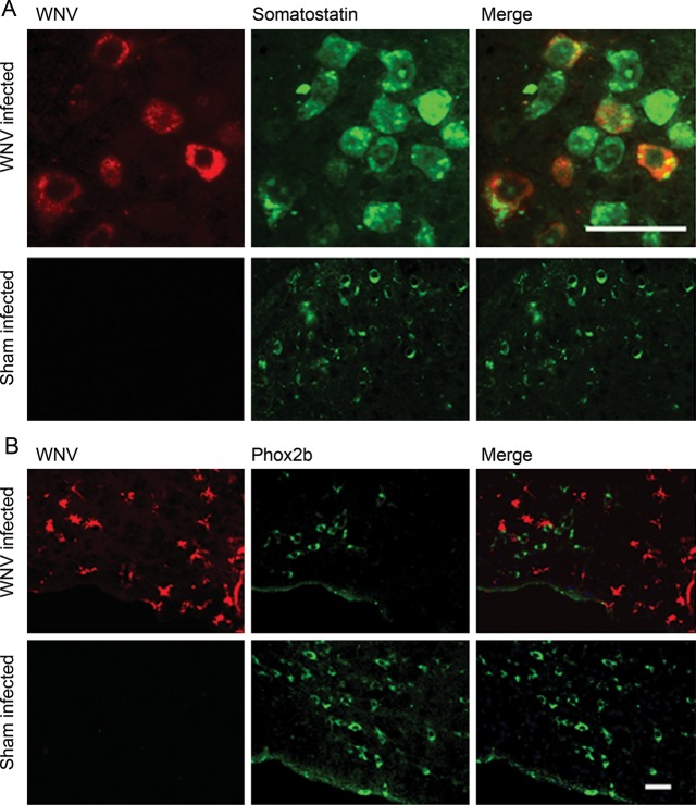
Results: Among infections due to all but 1 alphavirus, death was strongly associated with a suppressed minute volume. Virally infected mice with a very low minute volume did not neurologically respond to hypercapnia or optogenetic photoactivation of the C4 cervical cord. Neurons with the orexin 1 receptor protein in the ventral C3-5 cervical cord were statistically diminished in WNV-infected mice with a low minute volume as compared to WNV-infected or sham-infected mice without respiratory insufficiency. Also, WNV-infected cells were adjacent to neurons with respiratory functions in the medulla.
Conclusions: Detection of a common neurological mechanism of death among viral encephalitides creates opportunities to create broad-spectrum therapies that target relevant neurological cells in patients with types of viral encephalitis that have not been treatable in the past.
Keywords: West Nile virus; alphavirus; electromyography; encephalitis; flavivirus; neurons; optogenetic; respiratory.
Figures


Similar articles
-
Respiratory insufficiency correlated strongly with mortality of rodents infected with West Nile virus.PLoS One. 2012;7(6):e38672. doi: 10.1371/journal.pone.0038672. Epub 2012 Jun 14. PLoS One. 2012. PMID: 22719920 Free PMC article.
-
Neurological suppression of diaphragm electromyographs in hamsters infected with West Nile virus.J Neurovirol. 2010 Jul;16(4):318-29. doi: 10.3109/13550284.2010.501847. J Neurovirol. 2010. PMID: 20632796 Free PMC article.
-
Serological evidence of flaviviruses and alphaviruses in livestock and wildlife in Trinidad.Vector Borne Zoonotic Dis. 2012 Nov;12(11):969-78. doi: 10.1089/vbz.2012.0959. Epub 2012 Sep 18. Vector Borne Zoonotic Dis. 2012. PMID: 22989182 Free PMC article.
-
Astrocytes in Flavivirus Infections.Int J Mol Sci. 2019 Feb 6;20(3):691. doi: 10.3390/ijms20030691. Int J Mol Sci. 2019. PMID: 30736273 Free PMC article. Review.
-
Lipids and flaviviruses, present and future perspectives for the control of dengue, Zika, and West Nile viruses.Prog Lipid Res. 2016 Oct;64:123-137. doi: 10.1016/j.plipres.2016.09.005. Epub 2016 Oct 1. Prog Lipid Res. 2016. PMID: 27702593 Review.
Cited by
-
Autonomic deficit not the cause of death in West Nile virus neurological disease.Clin Auton Res. 2014 Feb;24(1):15-23. doi: 10.1007/s10286-013-0213-y. Epub 2013 Oct 25. Clin Auton Res. 2014. PMID: 24158383 Free PMC article.
-
Neurological approaches for investigating West Nile virus disease and its treatment in rodents.Antiviral Res. 2013 Nov;100(2):535-45. doi: 10.1016/j.antiviral.2013.09.010. Epub 2013 Sep 19. Antiviral Res. 2013. PMID: 24055448 Free PMC article. Review.
-
Phrenic nerve deficits and neurological immunopathology associated with acute West Nile virus infection in mice and hamsters.J Neurovirol. 2017 Apr;23(2):186-204. doi: 10.1007/s13365-016-0488-6. Epub 2016 Oct 19. J Neurovirol. 2017. PMID: 27761801 Free PMC article.
-
A case report of long-latency evoked diaphragm potentials after exposure to acute intermittent hypoxia in post-West Nile virus meningoencephalitis.J Neurophysiol. 2025 Feb 1;133(2):522-529. doi: 10.1152/jn.00406.2024. Epub 2024 Dec 30. J Neurophysiol. 2025. PMID: 39852952 Free PMC article.
-
Emerging infections of the central nervous system.Curr Infect Dis Rep. 2013 Dec;15(6):576-82. doi: 10.1007/s11908-013-0377-6. Curr Infect Dis Rep. 2013. PMID: 24136412
References
-
- Misra UK, Kalita J. Seizures in Japanese encephalitis. J Neurol Sci. 2001;190:57–60. - PubMed
-
- Paessler S, Aguilar P, Anishchenko M, et al. The hamster as an animal model for eastern equine encephalitis—and its use in studies of virus entrance into the brain. J Infect Dis. 2004;189:2072–6. - PubMed
-
- Kitchener N. Cerebral edema. In: Kitchener N, Hashem S, Wahba M, Khalaf M, Zarif B, Mansoor S, editors. Critical care in neurology. Wuppertal, Germany: Flying Publisher; 2012. pp. 79–83.
-
- Bannister R, Mathias CJ. Clinical features and evaluation of the primary chronic autonomic failure syndromes. In: Bannister R, Mathias CJ, editors. Autonomic failure: a textbook of clinical disorders of the autonomic nervous system. 4th ed. New York: Oxford University Press; 2004. pp. 307–16.
-
- Shindarov LM, Chumakov MP, Voroshilova MK, et al. Epidemiological, clinical, and pathomorphological characteristics of epidemic poliomyelitis-like disease caused by enterovirus 71. J Hyg Epidemiol Microbiol Immunol. 1979;23:284–95. - PubMed
Publication types
MeSH terms
Grants and funding
LinkOut - more resources
Full Text Sources
Other Literature Sources
Medical
Miscellaneous
