. 2013 Jul 15;126(Pt 14):3204-13.
doi: 10.1242/jcs.128587. Epub 2013 May 2.

Germline deletion of Cetn1 causes infertility in male mice

Affiliations

Germline deletion of Cetn1 causes infertility in male mice

Prachee Avasthi et al. J Cell Sci. .

Abstract

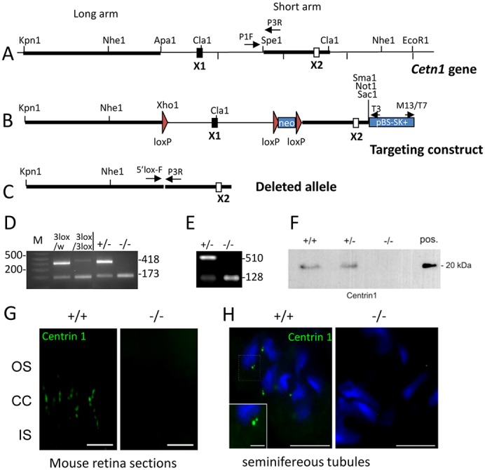
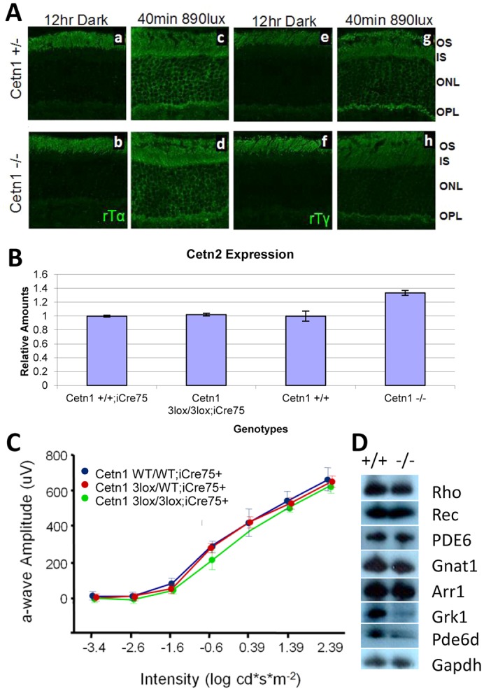
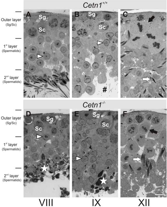
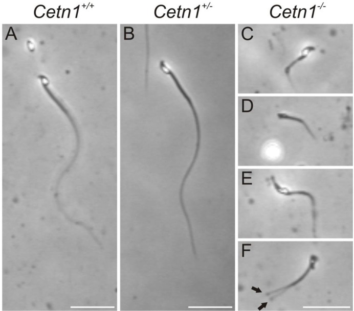
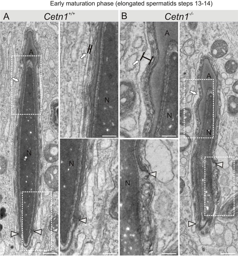
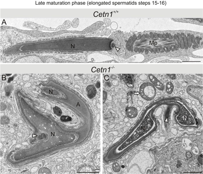
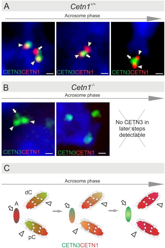
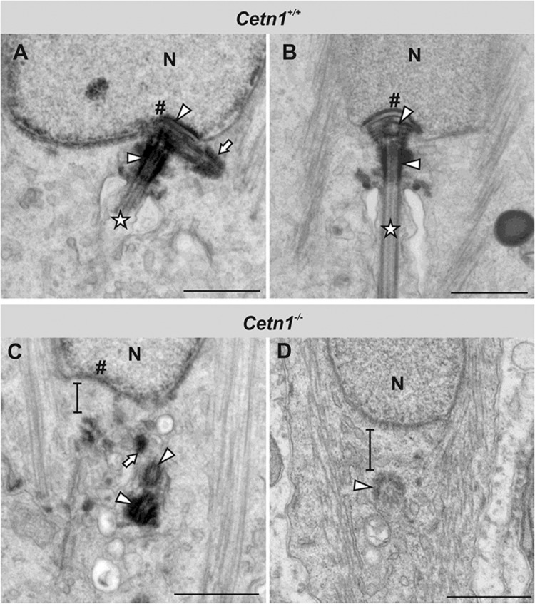
Centrins are calmodulin-like Ca(2+)-binding proteins that can be found in all ciliated eukaryotic cells from yeast to mammals. Expressed in male germ cells and photoreceptors, centrin 1 (CETN1) resides in the photoreceptor transition zone and connecting cilium. To identify its function in mammals, we deleted Cetn1 by homologous recombination. Cetn1(-/-) mice were viable and showed no sign of retina degeneration suggesting that CETN1 is nonessential for photoreceptor ciliogenesis or structural maintenance. Phototransduction components localized normally to the Cetn1(-/-) photoreceptor outer segments, and loss of CETN1 had no effect on light-induced translocation of transducin to the inner segment. Although Cetn1(-/-) females and Cetn1(+/-) males had normal fertility, Cetn1(-/-) males were infertile. The Cetn1(-/-) testes size was normal, and spermatogonia as well as spermatocytes developed normally. However, spermatids lacked tails suggesting severe defects at the late maturation phase of spermiogenesis. Viable sperm cells were absent and the few surviving spermatozoa were malformed. Light and electron microscopy analyses of Cetn1(-/-) spermatids revealed failures in centriole rearrangement during basal body maturation and in the basal-body-nucleus connection. These results confirm an essential role for CETN1 in late steps of spermiogenesis and spermatid maturation.

Keywords: CETN1 deletion; Centrin; Flagella; Photoreceptors; Spermatid maturation; Spermiogenesis.


Figures

Fig. 1.
Fig. 1.
Cetn1 deletion. (A) The endogenous Cetn1 locus with one coding exon (X1) and one non-coding exon (X2). Long arm, short arm, loxP sites (red triangles) and relevant enzyme restriction sites are indicated. (B) The 3loxP knock-in construct with loxP sites flanking exon1 and neo for eventual deletion. (C) The deleted Cetn1 gene. (D) Genotyping of Cetn1loxP/WT;iCre75+ (labeled 3lox/w) and of Cetn13loxP/3loxP;iCre75+ mice (labeled 3lox/3lox), and Cetn1+/− and Cetn1−/− mice with retina DNA as PCR template. PCR amplification detects a wild-type allele at 418 bp in Cetn1+/− and a deletion product at 173 bp in Cetn1−/− mice. (E) RT-PCR was performed on extracts from Cetn1+/− and Cetn1−/− testes; 510 bp amplicons were detected for Cetn1+/− but not for Cetn1−/−. (F) Immunoblot of wild-type and Cetn1−/− testes protein extract. CETN1 is undetectable in the germline knockout mouse. (G) Immunohistochemistry of wild-type and Cetn1−/− retina cryosections using the anti-CETN1 antibody, MmC1. CETN1 localizes to wild-type photoreceptor cilia, but is undetectable in the Cetn1−/− retina. Scale bar: 10 µm. (H) CETN1 immunohistochemistry of wild-type seminifereous tubules. Antibody MmC1 detects characteristic dots (centrioles), one of which is brighter and bigger at the condensed nuclei in Cetn1+/+tubules, whereas in Cetn1−/− tubules, labeling is undetectable. Scale bars: 10 µm; inset, 5 µm.
Fig. 2.
Fig. 2.
Normal transducin translocation and phototransduction in Cetn1−/− photoreceptors. (A) Light-induced translocation of transducin subunits, Tα and Tβγ. Cetn1+/− and Cetn1−/− mice were dark-adapted for 12 hours, or dark-adapted and exposed to light for 40 minutes. Retina cryosections were probed with anti-transducin antibodies, anti-Gnat1(Tα) and anti-Gngt1 (Tγ). Light-induced translocation of Tα and Tγ was indistinguishable in the two genotypes. (B) Upregulation of CETN2 in Cetn1−/− knockout retinas. cDNA from Cetn1WT/WT;iCre75+, Cetn13lox/3lox;iCre75+, Cetn1+/+, and Cetn1−/− retinas was amplified under real-time conditions (see Materials and Methods). No significant difference in Cetn2 expression was detected in Cetn13lox/3lox;iCre75+ (N = 2) compared with WT retinas. In global knockouts, Cetn2 was upregulated over 30% (N = 2) suggesting compensation for loss of the CETN1 isoform. (C) Electroretinogram of wild-type, heterozygous and homozygous Cetn1 rod-specific deletion retinas. (D) Immunoblots of outer segment polypeptides unaffected (rhodopsin, recoverin, PDE6, rod Tα, and rod arrestin), and downregulated (GRK1 and PDE6D) by Cetn1 deletion.
Fig. 3.
Fig. 3.
Seminifereous tubules of Cetn1+/+ and Cetn1−/− mice. Stage-matched seminifereous tubules from (A–C) Cetn1+/+ mice and (D–F) Cetn1−/− mice. The outer layer consists of spermatogonia (Sg) and spermatocytes (Sc), the first layer consists of spermatids (arrowheads) from round Golgi phase to the end of acrosome phase; the second layer harbors the maturating spermatids up to spermiation into the lumen of seminifereous tubules. (A) Stage VIII tubules of Cetn1+/+ mice with spermatocytes in the outer layer, round spermatids in the first layer and tailed spermatids in the second layer at the luminal side of the tubules. (B) Stage IX tubules of Cetn1+/+ mice. Instead of round spermatids, capped spermatids entering acrosome phase are present in the first tubule layer. #, Tails of mature spermatids after spermiation in the tubule lumen. (C) Stage XII tubules of Cetn1+/+ mice exhibit spermatocytes at meiosis (black arrow) in the first layer forming the new generation of spermatids. Elongated spermatids (white arrow) are present in the second layer. (D) Stage VIII tubules of Cetn1−/− mice with spermatocytes in the outer layer, and round spermatids in the first layer. Tailed spermatids in the second layer are absent, but dark, dense material of degraded spermatids (white star) is present. (E) Stage IX tubules of Cetn1−/− mice reveal the same layering as Cetn1+/+ mice, but contain dark dense material of degraded spermatids (white star). (F) Stage XII tubules of Cetn1−/− mice show spermatocytes during meiosis (black arrow) in the first layer and elongated spermatids at the transition to maturation phase (white arrow). Layering of the seminifereous tubules was the same in Cetn1+/+ and Cetn1−/− mice. Scale bar: 10 µm.
Fig. 4.
Fig. 4.
Light microscopy analysis of sperm present in spermatic duct. Equal amounts of sperm from Cetn1+/+, Cetn1+/− and Cetn1−/− mice were collected and analyzed by light microscopy. (A,F) Sperm extracted from Cetn1+/+ and Cetn1+/− mice were identical in shape and length. (B–E) Sperm from Cetn1−/− mice were all malformed in one or more ways: sperm tail shape, sperm tail length, head shape and midpiece. Bifurcated tail-like structures (F, arrows) were sometimes observed. Scale bars: 25 µm.
Fig. 5.
Fig. 5.
Ultrastructure of early maturation phase spermiogenesis of Cetn1+/+ and Cetn1−/− spermatids. (A,B) The first differences between Cetn1+/+ and Cetn1−/− were found in steps 13–14. The caudal movement of the acrosome forming the acrosomic clefts (arrowheads) was uneven and a general bulky appearance was observed in Cetn1−/− spermatids. Sertoli cell membranes were deformed compared to the straight, parallel membranes of Cetn1+/+ mice (arrows). At higher magnification (center two panels), wider distention between the Sertoli cell membranes was seen (arrows, black bars). Also at high magnification, at the spermatid base a disruption to the caudal migration of the acrosome was evident (B, arrowheads). Scale bars: 0.5 µm.
Fig. 6.
Fig. 6.
Late maturation phase spermatids. Spermatids were analyzed by high resolution electron microscopy at steps 15–16 of spermiogenesis. (A) Wild-type spermatid showing a midpiece (Mp), an elongated nucleus (N) and a remaining centriolar structure (arrowhead). (B,C) Abnormalities were prominent in Cetn1−/− spermatids. No midpiece or tail formation was found. Nuclei were split, bifurcated and the chromatin appeared perforated. Additionally, spermatid heads were bent over and the shape of the centrosomal region (arrowheads) was abnormal. Cell debris of spermatids in granules (C, arrow) was observed, indicating cell degeneration. A, acrosome. Scale bars: 1 µm.
Fig. 7.
Fig. 7.
CETN1 expression during centriole rearrangement in spermatids in late spermiogenesis. (A,B) Centrin expression was detected by immunofluorescence. Double-labeling of CETN1 (red) and the centriole marker CETN3 (green) in acrosome phase spermatids (steps 8–12) of Cetn1+/+ and Cetn1−/− mice. (A) CETN1 and CETN3 were colocalized in the centrioles of spermatid centrosomes, the distal and the proximal centrioles (arrows) of the sperm flagellar basal apparatus as well as the centriolar adjunct (arrowheads) in Cetn1+/+ mice. (B) CETN1 was absent, centriole arrangement of the flagellar basal apparatus was disorganized and spermatogenesis was arrested in the last phase in Cetn1−/− mice. No CETN3 staining was identified in later steps of spermiogenesis (indicated by the X). (C) Scheme representing the polarization of CETN3/CETN1 during the acrosome phase; A, adjunct; dC, distal centriole; pC, proximal centriole; arrows and arrowheads as in A. Scale bars: 0.5 µm.
Fig. 8.
Fig. 8.
Ultrastructure of spermatid centriole rearrangement during late spermiogenesis. (A,B) Cetn1+/+ spermatids during centriole rearrangement of late spermiogenesis. (A) Proximal and distal centrioles (arrowheads) positioned directly adjacent to the fossa (#) of the nucleus (N). The proximal centriole projects the adjunct (arrow), and the sperm flagellum axoneme is visible (star). (B) Rearranged centrioles (arrowheads) finish to form the basal body apparatus at the nuclear fossa (#), from which the sperm flagellar axoneme extends (star). (C,D) Cetn1−/− spermatids during centriole rearrangement. (C) Both centrioles (arrowheads) and the adjunct remnant (arrow) appear disengaged from the nuclear membrane, as indicated by the black bar. The nuclear fossa (#) is dysmorphic. (D) The distal centriole (arrowhead) projects a tubular structure corresponding to the axoneme, but it is bent and flaccid. Instead of a modified proximal centriole, a cytoplasmic gap (indicated by black bar) appears between the nucleus (N) and the distal centriole. Scale bars: 0.5 µm.

References

    1. Avasthi P., Watt C. B., Williams D. S., Le Y. Z., Li S., Chen C. K., Marc R. E., Frederick J. M., Baehr W. (2009). Trafficking of membrane proteins to cone but not rod outer segments is dependent on heterotrimeric kinesin-II. J. Neurosci. 29, 14287–14298 10.1523/JNEUROSCI.3976-09.2009 - DOI - PMC - PubMed
    1. Baehr W., Karan S., Maeda T., Luo D. G., Li S., Bronson J. D., Watt C. B., Yau K. W., Frederick J. M., Palczewski K. (2007). The function of guanylate cyclase 1 and guanylate cyclase 2 in rod and cone photoreceptors. J. Biol. Chem. 282, 8837–8847 10.1074/jbc.M610369200 - DOI - PMC - PubMed
    1. Brugerolle G., Mignot J. P. (2003). The rhizoplast of chrysomonads, a basal body-nucleus connector that polarises the dividing spindle. Protoplasma 222, 13–21 10.1007/s00709-003-0016-4 - DOI - PubMed
    1. Calvert P. D., Strissel K. J., Schiesser W. E., Pugh E. N., Jr, Arshavsky V. Y. (2006). Light-driven translocation of signaling proteins in vertebrate photoreceptors. Trends Cell Biol. 16, 560–568 10.1016/j.tcb.2006.09.001 - DOI - PubMed
    1. Cooke H. J., Saunders P. T. (2002). Mouse models of male infertility. Nat. Rev. Genet. 3, 790–801 10.1038/nrg911 - DOI - PubMed

Publication types

MeSH terms