. 2013 Jul 15;126(Pt 14):3070-81.
doi: 10.1242/jcs.122184. Epub 2013 May 2.

Direct binding of TUBB3 with DCC couples netrin-1 signaling to intracellular microtubule dynamics in axon outgrowth and guidance

Affiliations

Direct binding of TUBB3 with DCC couples netrin-1 signaling to intracellular microtubule dynamics in axon outgrowth and guidance

Chao Qu et al. J Cell Sci. .

Abstract

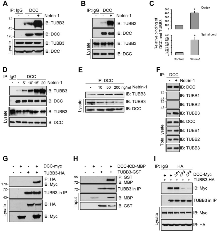
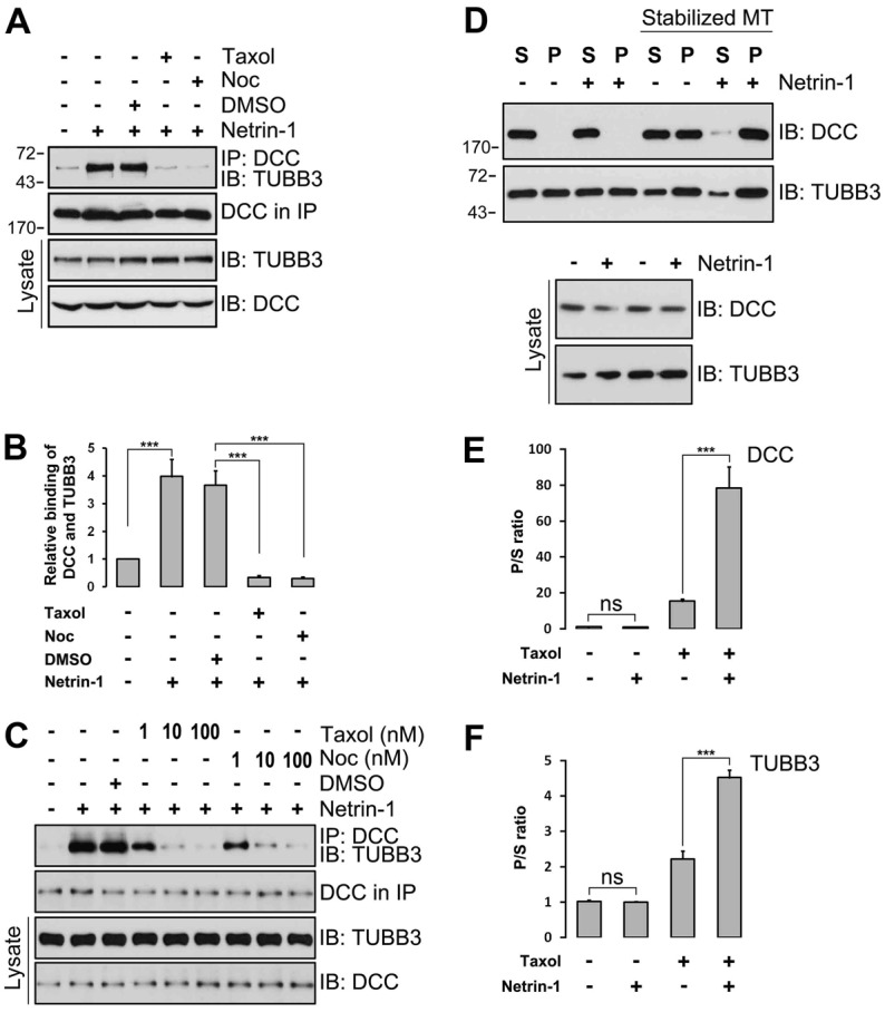
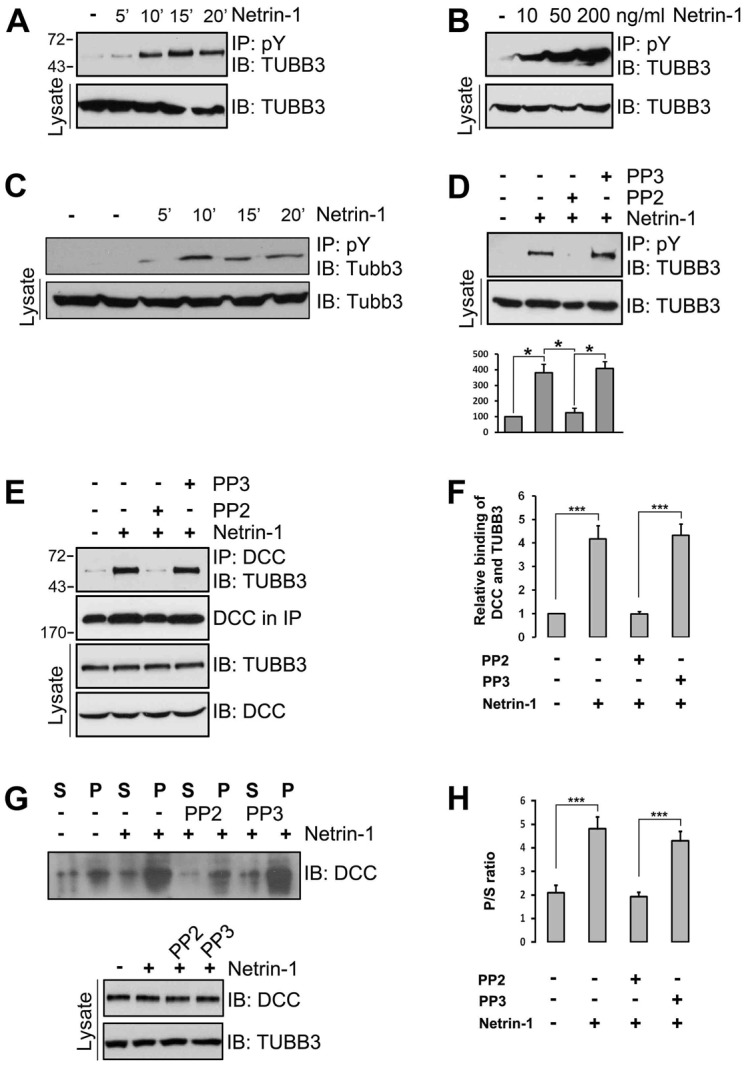
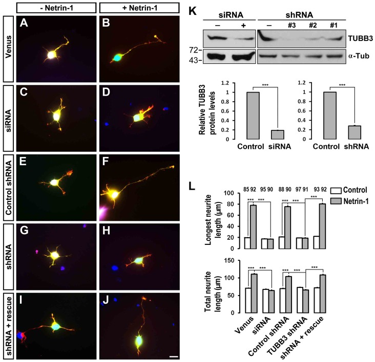
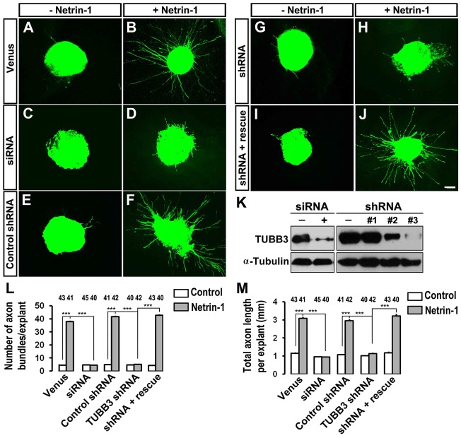
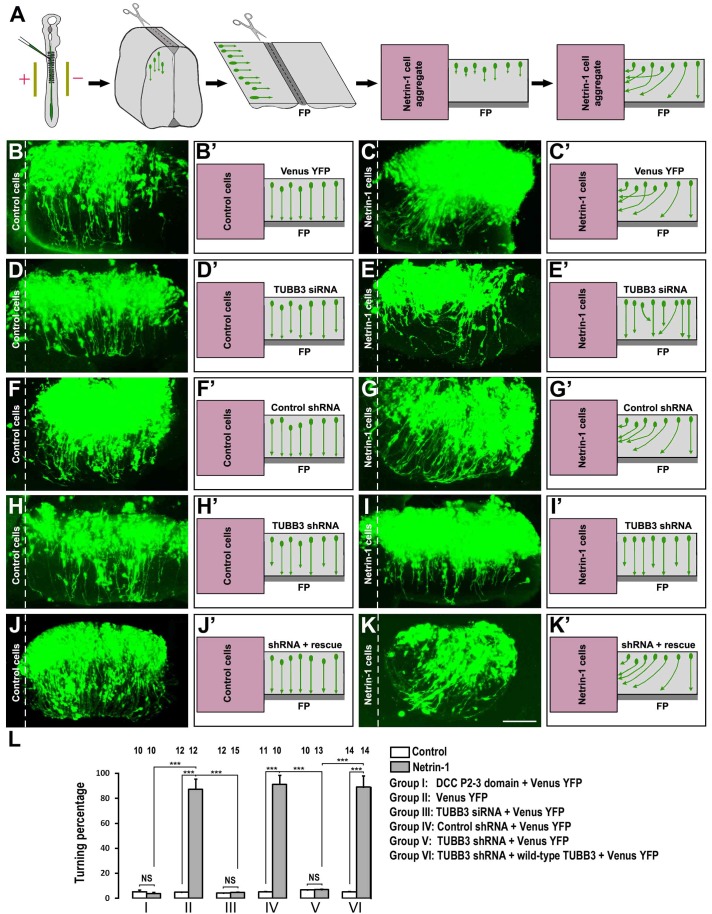
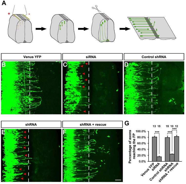
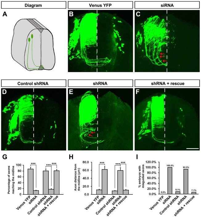
The coupling of axon guidance cues, such as netrin-1, to microtubule (MT) dynamics is essential for growth cone navigation in the developing nervous system. However, whether axon guidance signaling regulates MT dynamics directly or indirectly is unclear. Here, we report that TUBB3, the most dynamic β-tubulin isoform in neurons, directly interacts with the netrin receptor DCC, and that netrin-1 induces this interaction in primary neurons. TUBB3 colocalizes with DCC in the growth cones of primary neurons and MT dynamics is required for netrin-1-promoted association of TUBB3 with DCC. Netrin-1 not only increases co-sedimentation of DCC with polymerized MT, but also promotes MT dynamics in the growth cone. Knocking down TUBB3 inhibits netrin-1-induced MT dynamics, axon outgrowth and attraction in vitro and causes defects in commissural axon projection in the embryo. These results indicate that TUBB3 directly links netrin signaling pathways to MT dynamics and plays an important role in guiding commissural axons in vivo.

Keywords: Axon guidance; DCC; Microtubule dynamics; Netrin; Signal transduction; TUBB3.


Figures

Fig. 1.
Fig. 1.
Interaction of TUBB3 with DCC. (A,B) Interaction of endogenous TUBB3 with DCC in E15 cortical (A) and E13 dorsal spinal cord (B) neurons. (C) Quantification of A and B from three independent experiments; *P<0.05 (two-tailed Student's t-test). (D) Netrin-1 increased the interaction of endogenous TUBB3 with DCC in a time-dependent manner. Lysates of dissociated neurons from E13 mouse spinal cords were immunoprecipitated with anti-DCC and analyzed with anti-TUBB3. (E) Netrin-1 increased the interaction of endogenous TUBB3 with DCC in a dose-dependent manner. E15 primary cortical neurons were treated with purified netrin-1 at 10, 50 and 200 ng/ml. (F) Endogenous DCC interacted with TUBB3, not TUBB1 and TUBB2, in E15 cortical neurons with or without netrin-1 treatment. Cell lysates of dissociated neurons from E15 mouse cortexes were immunoprecipitated with anti-DCC and followed by probing with anti-TUBB1, anti-TUBB2 or anti-TUBB3. (G) Interaction of TUBB3 with DCC in HEK293 cells. TUBB3-HA was co-transfected with DCC-Myc into HEK293 cells. Anti-HA (TUBB3) precipitated DCC-Myc. (H) Direct interaction of TUBB3 with DCC. Purified TUBB3 was incubated with purified intracellular domain of DCC tagged with MBP in vitro. The anti-GST antibody was used to immunoprecipitate proteins and the blot was analyzed with anti-MBP. (I) P2 and P3 domains in DCC are required for the interaction of DCC and TUBB3. TUBB3-HA was co-transfected with different truncated DCCs (DCC-ΔP1, ΔP2 and ΔP3) tagged with Myc in HEK293 cells.
Fig. 2.
Fig. 2.
The induction of the interaction of TUBB3 with DCC by netrin-1 depends on MT dynamics. (AC) Taxol and nocodazole (Noc) inhibited netrin-1-induced interactions of endogenous TUBB3 with DCC. E15 cortical neurons were treated with purified netrin-1 in the presence of different concentrations of taxol or nocodazole (1 µM taxol and 3 µM nocodazole in A). (B) Quantification of A from three independent experiments showing relative binding of DCC and TUBB3. (DF) E15 cortical neurons were stimulated with netrin-1 and the co-sedimentation assay of cell lysates was performed in the absence or presence of taxol. DCC and TUBB3 in the pellet (P) and supernatant (S) fractions were examined by western blot using anti-DCC and anti-TUBB3, respectively. (E,F) Quantification of three independent experiments showing P/S ratio of DCC (E) and TUBB3 (F), respectively. Netrin-1 increased the co-sedimentation of DCC and TUBB3 with polymerized MTs in primary neurons. ns, not significant; ***P<0.001 (two-tailed Student's t-test).
Fig. 3.
Fig. 3.
Src family kinase activity is required for the induction of TUBB3 tyrosine phosphorylation and the interaction of TUBB3 with DCC by netrin-1. (A,B) Induction of TUBB3 tyrosine phosphorylation in dissociated cortical neurons by netrin-1. E15 cortical neurons were treated with purified netrin-1 for 5–20 minutes. The anti-pY antibody was used to immunoprecipitate proteins and the immunoblot was analyzed with anti-TUBB3. (C) Induction of TUBB3 tyrosine phosphorylation in dissociated E13 dorsal spinal cord neurons by netrin-1. Primary neurons were treated with purified netrin-1 (200 ng/ml). (D) Src family kinase-specific inhibitor PP2 inhibited netrin-1-induced TUBB3 tyrosine phosphorylation. E15 cortical cells were stimulated with netrin-1 in the presence of PP2 or PP3. Quantification is shown in the lower panel (Student's t-test). (E) PP2, but not PP3, blocked netrin-1-induced interaction of endogenous TUBB3 with DCC in primary neurons. (F) Quantification showing relative binding of DCC and TUBB3 in E. (G,H) Netrin-1-stimulated co-sedimentation of DCC with MTs was inhibited by PP2, not PP3. E15 cortical neurons were stimulated with netrin-1 in the presence of PP2 or PP3. DCC in the pellet and supernatant fractions was examined by western blot. Quantification of G is shown in H (one-way ANOVA and Fisher LSD post-hoc comparisons). *P<0.05, ***P<0.001.
Fig. 4.
Fig. 4.
Inhibition of netrin-1-induced axon outgrowth of cortical neurons by TUBB3 knockdown. (AJ) E15 mouse cortical neurons were transfected with Venus YFP only (A,B), Venus YFP plus the TUBB3 siRNA pool (C,D), Venus YFP plus control shRNA (E,F), Venus YFP plus TUBB3 shRNA (G,H) and Venus YFP plus shRNA and the wild-type human RNAi-resistant TUBB3 (I,J). Neurite outgrowth from YFP-positive neurons was assessed in the presence of purified netrin-1 (B,D,F,H,J) and in the sham-purified control (A,C,E,G,I). (K) Both TUBB3 siRNA pool and shRNA (#3) reduced endogenous TUBB3 protein levels in E15 cortical neurons (Student's t-test). (L) Quantification of netrin-1-induced neurite outgrowth. Only the neurites of YFP-positive neurons not in contact with other cells were measured and used in the statistical analyses. Data are mean ± s.e.m. from three separate experiments. The numbers on the top of each bar indicate the numbers of neurons tested in the corresponding groups (one-way ANOVA with Fischer LSD for post-hoc comparisons). ***P<0.001. Scale bar: 10 µm.
Fig. 5.
Fig. 5.
Inhibition of netrin-1-induced axon outgrowth of chick dorsal spinal cord explants by TUBB3 knockdown. (AJ) Chick neural tubes were electroporated in ovo at stage 12–15 with Venus YFP only (A,B), Venus YFP plus the TUBB3 siRNA pool (C,D), Venus YFP plus control shRNA (E,F), Venus YFP plus TUBB3 shRNA (G,H) and Venus YFP plus TUBB3 shRNA and the wild-type human RNAi-resistant TUBB3 (I,J). Netrin-1 increased commissural axon outgrowth with longer and more axon bundles in the netrin-1 group (B,F) than the control group (A,E). Netrin-1-induced axon outgrowth was inhibited either by the TUBB3 siRNA pool (C,D) or shRNA (G,H). The expression of the wild-type human RNAi-resistant TUBB3 plasmids reversed the effect of TUBB3 shRNA (I,J). (K) Both TUBB3 siRNA pool and shRNA efficiently knocked down endogenous TUBB3 in chick spinal cords. (L,M) Quantification of netrin-1-induced commissural axon outgrowth. Only YFP-positive axon bundles were measured and used in the statistical analyses. The numbers on the top of each bar indicate the numbers of explants tested in the corresponding groups. Data are mean ± s.e.m. from three separate experiments. ***P<0.001 (one-way ANOVA with Fischer LSD for post-hoc comparisons). Scale bar: 100 µm.
Fig. 6.
Fig. 6.
Requirement of TUBB3 for netrin-1 attraction of spinal commissural axons. (A) Diagram of the in ovo electroporation and the co-culture assay with the open-book preparation. (BK′) Electroporation of Venus YFP into the neural tube of chick embryos allowed visualization of axons. In the left panels (B,B′,D,D′,F,F′,H,H′,J,J′), neural tube explants were co-cultured with control HEK293 cells and commissural axons projected straight toward the floor plate (FP). In the right panels (C,C′,E,E′,G,G′,I,I′,K,K′), neural tube explants were co-cultured with aggregates of HEK293 cells secreting netrin-1. The neural tube was electroporated with Venus YFP alone (B,B′,C,C′); Venus YFP together with the TUBB3 siRNA pool (D,D′,E,E′); Venus YFP together with the control shRNA (F,F′,G,G′); Venus YFP with the TUBB3 shRNA (H,H′,I,I′) and Venus YFP with the TUBB3 shRNA plus the wild-type human RNAi-resistant TUBB3 (J,J′,K,K′). Expression of either TUBB3 siRNAs or shRNA inhibited commissural axon turning towards the netrin-1 source. The commissural axon turning defect of RNAi knockdown could be rescued by expressing RNAi-resistant TUBB3. (L) Quantification of axon turning. The numbers on the top of each bar indicate the numbers of explants tested in the corresponding groups. Data are mean ± s.e.m. from groups I–VI. ***P<0.001 (one-way ANOVA and Fisher LSD post-hoc comparisons). NS, not significant. Scale bar:100 µm.
Fig. 7.
Fig. 7.
Inhibition of commissural axon projection in vivo by TUBB3 RNAi. (A) Diagram showing the experimental design. (BF) Different combinations of plasmids and siRNAs were electroporated into the chick neural tube in ovo at stage 12 and the lumbosacral region of the spinal cord was isolated at stage 23. (B) Neurons electroporated with Venus YFP only. (C) Neurons electroporated with Venus YFP plus the TUBB3 siRNA pool. (D) Neurons with Venus YFP plus control shRNA. (E) Neurons with Venus YFP plus TUBB3 shRNA. (F) Neurons with Venus YFP plus shRNA and wild-type human RNAi-resistant TUBB3. The red arrowheads point to shortened axons. (G) Quantification of the percentage of axons reaching the FP. The numbers on the top of each bar indicate the numbers of embryos tested in the corresponding groups. ***P<0.001 (one-way ANOVA with Fischer LSD for post-hoc comparisons). Scale bar: 100 µm.
Fig. 8.
Fig. 8.
TUBB3 is essential for spinal cord commissural axon pathfinding in vivo. (A) Diagram showing the transverse section of the chick spinal cord after electroporation. (BF) The chick neural tube was electroporated with Venus YFP only (B), Venus YFP plus the TUBB3 siRNA pool (C), Venus YFP plus control shRNA (D), Venus YFP plus TUBB3 shRNA (E) or Venus YFP plus shRNA and wild-type human RNAi-resistant TUBB3 (F). Expression of TUBB3 siRNAs or shRNA not only inhibited the commissural axon extension but also caused aberrant pathfinding (C,E). The wild-type RNAi-resistant TUBB3 rescued the defect of TUBB3 shRNA knockdown on commissural axon projection and turning (F). The red arrows point to misguided axons. (G) Quantification of the percentage of axons reaching the midline of the chick spinal cord. (H) Quantification of the average distance of axons away from the midline. (I) The percentage of embryos with misguided axons. The numbers of embryos tested were 17 for the Venus YFP group, 14 for the siRNA pool group, 12 for the control shRNA group, 15 for the TUBB3 shRNA group and 14 for the rescue group. ***P<0.001 (one-way ANOVA with Fischer LSD for post-hoc comparisons). Scale bar: 100 µm.

Similar articles

Cited by

References

    1. Ackerman S. L., Kozak L. P., Przyborski S. A., Rund L. A., Boyer B. B., Knowles B. B. (1997). The mouse rostral cerebellar malformation gene encodes an UNC-5-like protein. Nature 386, 838–842 10.1038/386838a0 - DOI - PubMed
    1. Alcántara S., Ruiz M., De Castro F., Soriano E., Sotelo C. (2000). Netrin 1 acts as an attractive or as a repulsive cue for distinct migrating neurons during the development of the cerebellar system. Development 127, 1359–1372 - PubMed
    1. Buck K. B., Zheng J. Q. (2002). Growth cone turning induced by direct local modification of microtubule dynamics. J. Neurosci. 22, 9358–9367 - PMC - PubMed
    1. Colamarino S. A., Tessier-Lavigne M. (1995). The axonal chemoattractant netrin-1 is also a chemorepellent for trochlear motor axons. Cell 81, 621–629 10.1016/0092-8674(95)90083-7 - DOI - PubMed
    1. Dent E. W., Gertler F. B. (2003). Cytoskeletal dynamics and transport in growth cone motility and axon guidance. Neuron 40, 209–227 10.1016/S0896-6273(03)00633-0 - DOI - PubMed

Publication types

MeSH terms