Morphological spectrum of pilar cysts
- PMID: 23641374
- PMCID: PMC3624713
- DOI: 10.4103/1947-2714.107532
Morphological spectrum of pilar cysts
Abstract
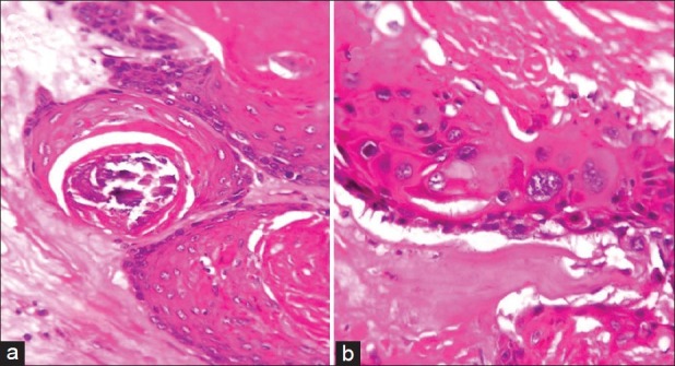
Background: Cysts of the skin are one of the commonly excised specimens in the surgical outpatient department. A majority of them being clinically diagnosed as sebaceous cysts, their true nature is only discernible on histopathological examination. Closer examination of the type of keratinization involved will throw light into the exact nature of the cyst. Trichilemmal or Pilar cyst is one such entity, which presents in both a non-neoplastic and neoplastic form.
Aims: The present retrospective observational study was undertaken to find out the incidence of these cysts in surgical pathology practice in a rural hospital and to enlist the various morphological forms that these cysts may take.
Materials and methods: The histopathology files were reviewed for a period of 6 years for cases coded as pilar cyst.
Results: A total of eight cases (5.75%) were identified, which showed features of trichilemmal differentiation. A single case each of proliferating trichilemmal cyst and malignant proliferating trichilemmal tumors were noted. Most of the cases were seen among females on the scalp.
Conclusions: Trichilemmal tumor is an uncommon histopathological entity. Many of these lesions may be mistakenly diagnosed due to lack of recognition of the unique type of keratinization.
Keywords: Malignant pilar tumor; Pilarcyst; Proliferating pilar tumor; Proliferating trichilemmal cyst.
Conflict of interest statement
Figures


Similar articles
-
Cytodiagnosis of simple and proliferating trichilemmal cysts.Acta Cytol. 2001 Jul-Aug;45(4):582-8. doi: 10.1159/000327868. Acta Cytol. 2001. PMID: 11480722
-
A Proliferating Trichilemmal Tumor at an Uncommon Site Treated With Radical Excision: A Case Report and Literature Review.Cureus. 2024 Jul 18;16(7):e64803. doi: 10.7759/cureus.64803. eCollection 2024 Jul. Cureus. 2024. PMID: 39156394 Free PMC article.
-
Laryngeal Pilar Cyst Masquerading as an Internal/External Laryngocele.Clin Med Insights Ear Nose Throat. 2018 Dec 4;11:1179550618815917. doi: 10.1177/1179550618815917. eCollection 2018. Clin Med Insights Ear Nose Throat. 2018. PMID: 30574000 Free PMC article.
-
Two Cases of Malignant Proliferating Trichilemmal Tumor (MPTT) and Review of Literature.R I Med J (2013). 2022 Feb 1;105(1):12-16. R I Med J (2013). 2022. PMID: 35081182 Review.
-
Proliferating Pilar Tumor: Two Cases and a Review of the Literature.J Drugs Dermatol. 2021 Dec 1;20(12):1346-1348. doi: 10.36849/jdd.5978. J Drugs Dermatol. 2021. PMID: 34898151 Review.
Cited by
-
Scrotal trichilemmal cysts: a case report.Ann Med Surg (Lond). 2023 Apr 14;85(5):2166-2168. doi: 10.1097/MS9.0000000000000666. eCollection 2023 May. Ann Med Surg (Lond). 2023. PMID: 37229016 Free PMC article.
-
Giant, Bleeding, and Ulcerating Proliferating Trichilemmal Cyst, With Delayed Treatment Due to Coronavirus Outbreak: A Case Report and Review of the Literature.Front Surg. 2021 Nov 26;8:680160. doi: 10.3389/fsurg.2021.680160. eCollection 2021. Front Surg. 2021. PMID: 34901133 Free PMC article.
-
Pilar Cyst From a Maxillofacial Surgeon's Perspective: A Case Report and Review of Literature.Cureus. 2023 May 3;15(5):e38508. doi: 10.7759/cureus.38508. eCollection 2023 May. Cureus. 2023. PMID: 37273408 Free PMC article.
-
Pilar Cyst of the Dorsal Hand: A Rare Presentation of a Common Cyst.Cureus. 2024 May 22;16(5):e60865. doi: 10.7759/cureus.60865. eCollection 2024 May. Cureus. 2024. PMID: 38910694 Free PMC article.
-
Imaging findings of trichilemmal cyst and proliferating trichilemmal tumour.Neuroradiol J. 2021 Dec;34(6):615-621. doi: 10.1177/19714009211017789. Epub 2021 Jun 1. Neuroradiol J. 2021. PMID: 34060944 Free PMC article.
References
-
- Kirkham N. Tumors and cysts of the epidermis. In: Elder DE, Elenitsas R, Johnson BL, Murphy GF, editors. Lever's Histopathology of the skin. 9th ed. Philadelphia: Lippincott Williams and Wilkins; 2005. pp. 814–6.
-
- Rosai J. Rosai and Ackerman's surgical pathology. 9th ed. Vol. 1. St Louis: Mosby; 2004. pp. 151–3.
-
- Weedon D. Skin pathology. 2nd ed. Edinburgh: Churchill Livingstone; 2002. pp. 504–7.
LinkOut - more resources
Full Text Sources
Other Literature Sources