

Simple method to enhance the photostability of the fluorescence reporter R6G for prolonged single-molecule studies
- PMID: 23641719
- PMCID: PMC3723744
- DOI: 10.1021/jp4003643
Simple method to enhance the photostability of the fluorescence reporter R6G for prolonged single-molecule studies
Abstract
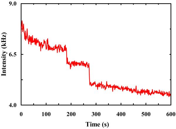
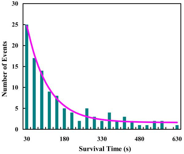
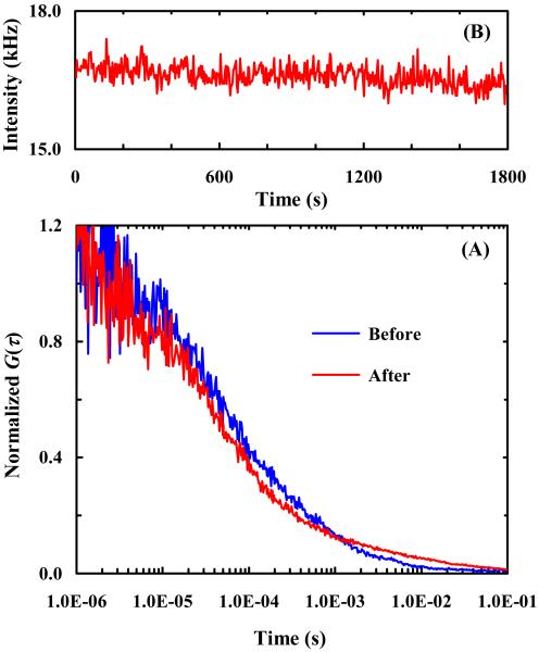
For fluorescence-based single-molecule studies, photobleaching of the dye reporter often limits the time window over which individual molecules can be followed. As such, many strategies, for example, using a cocktail of chemical reagents, have been developed to decrease the rate of photobleaching. Herein, we introduce a new and highly effective method to enhance the photostability of one of the commonly used fluorescent dyes, rhodamine 6G (R6G). We show that micrometer-sized polydimethylsiloxane (PDMS) wells, when the PDMS surface is properly treated, not only provide a confined environment for single-molecule detection but can also significantly increase the survival time of individual R6G molecules before photobleaching. Moreover, our results suggest, consistent with several previous studies, that R6G photobleaching involves a radical state.
Figures
Similar articles
-
Quantitative accounting of dye leakage and photobleaching in single lipid vesicle measurements: Implications for biomacromolecular interaction analysis.Colloids Surf B Biointerfaces. 2019 Oct 1;182:110338. doi: 10.1016/j.colsurfb.2019.06.067. Epub 2019 Jun 30. Colloids Surf B Biointerfaces. 2019. PMID: 31301580
-
Confeito-like assembly of organosilicate-caged fluorophores: ultrabright suprananoparticles for fluorescence imaging.Nanotechnology. 2012 May 4;23(17):175601. doi: 10.1088/0957-4484/23/17/175601. Epub 2012 Apr 5. Nanotechnology. 2012. PMID: 22481044 Free PMC article.
-
Molecular photobleaching kinetics of Rhodamine 6G by one- and two-photon induced confocal fluorescence microscopy.Chemphyschem. 2005 May;6(5):791-804. doi: 10.1002/cphc.200400509. Chemphyschem. 2005. PMID: 15884061 Review.
-
Micelle-vesicle-micelle transition in aqueous solution of anionic surfactant and cationic imidazolium surfactants: Alteration of the location of different fluorophores.J Colloid Interface Sci. 2017 Mar 15;490:762-773. doi: 10.1016/j.jcis.2016.12.009. Epub 2016 Dec 8. J Colloid Interface Sci. 2017. PMID: 27997846
-
Lumos maxima - How robust fluorophores resist photobleaching?Curr Opin Chem Biol. 2024 Apr;79:102439. doi: 10.1016/j.cbpa.2024.102439. Epub 2024 Mar 2. Curr Opin Chem Biol. 2024. PMID: 38432145 Review.
Cited by
-
Improved Dye Survival in Expansion Microscopy through Stabilizer-Conjugated Linkers.Chemistry. 2022 Nov 25;28(66):e202202404. doi: 10.1002/chem.202202404. Epub 2022 Sep 29. Chemistry. 2022. PMID: 36031562 Free PMC article.
References
-
- Joo C, Balci H, Ishitsuka Y, Buranachai C, Ha T. Advances in Single-Molecule Fluorescence Methods for Molecular Biology. Annu. Rev. Biochem. 2008;77:51–76. - PubMed
Publication types
MeSH terms
Substances
Grants and funding
LinkOut - more resources
Full Text Sources
Other Literature Sources
Research Materials
