. 2013 May 4;6(1):7.
doi: 10.1186/1756-6614-6-7.

A solitary hyperfunctioning thyroid nodule harboring thyroid carcinoma: review of the literature

Affiliations

A solitary hyperfunctioning thyroid nodule harboring thyroid carcinoma: review of the literature

Sasan Mirfakhraee et al. Thyroid Res. .

Abstract

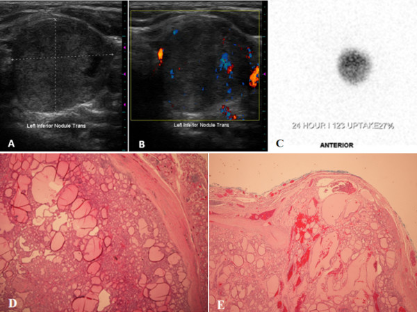
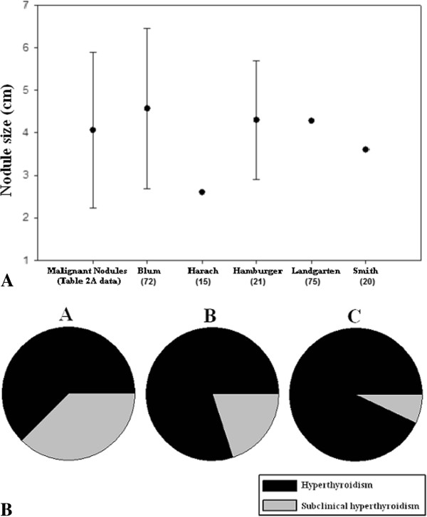
Hyperfunctioning nodules of the thyroid are thought to only rarely harbor thyroid cancer, and thus are infrequently biopsied. Here, we present the case of a patient with a hyperfunctioning thyroid nodule harboring thyroid carcinoma and, using MEDLINE literature searches, set out to determine the prevalence of and characteristics of malignant "hot" nodules as a group. Historical, biochemical and radiologic characteristics of the case subjects and their nodules were compared to those in cases of benign hyperfunctioning nodules. A literature review of surgical patients with solitary hyperfunctioning thyroid nodules managed by thyroid resection revealed an estimated 3.1% prevalence of malignancy. A separate literature search uncovered 76 cases of reported malignant hot thyroid nodules, besides the present case. Of these, 78% were female and mean age at time of diagnosis was 47 years. Mean nodule size was 4.13 ± 1.68 cm. Laboratory assessment revealed T3 elevation in 76.5%, T4 elevation in 51.9%, and subclinical hyperthyroidism in 13% of patients. Histological diagnosis was papillary thyroid carcinoma (PTC) in 57.1%, follicular thyroid carcinoma (FTC) in 36.4%, and Hurthle cell carcinoma in 7.8% of patients. Thus, hot thyroid nodules harbor a low but non-trivial rate of malignancy. Compared to individuals with benign hyperfunctioning thyroid nodules, those with malignant hyperfunctioning nodules are younger and more predominantly female. Also, FTC and Hurthle cell carcinoma are found more frequently in hot nodules than in general. We were unable to find any specific characteristics that could be used to distinguish between malignant and benign hot nodules.


Figures

Figure 1
Figure 1
Imaging and histologic features of the hot nodule present in the case report subject. (A) Ultrasonography of the left thyroid lobe, demonstrating a 2.7 cm, predominantly solid, and isoechoic nodule. (B) Color Doppler evaluation reveals blood flow within the rim of the nodule and intraparenchymally. (C) 123I thyroid scintigram depicts a round left-sided focus of iodine uptake with suppression in the remainder of the gland, consistent with an autonomously-functioning thyroid nodule. (D) Histological evaluation reveals that the lesion is solitary, circumscribed and encapsulated. The follicular proliferation is surrounded by a rather thick fibrous capsule. The lesion demonstrates a predominant follicular pattern of growth without papillary cytologic features (hematoxylin-eosin stain; original magnification × 4). (E) A focal area is identified where the tumor invades through and into the fibrous capsule (hematoxylin-eosin stain; original magnification × 2).
Figure 2
Figure 2
Size and biochemical assessment of hyperfunctioning thyroid nodules. (A) The mean greatest dimension of the malignant hot thyroid nodules from our case series is compared with that from five published surgical cases series of solitary, hyperfunctioning thyroid nodules. (B) The proportion of subjects with scintigraphically-determined hyperfunctioning thyroid carcinoma (Tables 2) who have frank biochemical hyperthyroidism vs. subclinical hyperthyroidism, based on varying nodule size. Subjects are characterized as having nodules < 2.5 cm (A), 2.5 – 4.5 cm (B), and > 4.5 cm (C) in diameter.

Similar articles

Cited by

References

    1. Vander JB, Gaston EA, Dawber TR. The significance of nontoxic thyroid nodules. Final report of a 15-year study of the incidence of thyroid malignancy. Ann Intern Med. 1968;69:537–540. doi: 10.7326/0003-4819-69-3-537. - DOI - PubMed
    1. Reiners C, Wegscheider K, Schicha H, Theissen P, Vaupel R, Wrbitzky R, Schumm-Draeger PM. Prevalence of thyroid disorders in the working population of Germany: ultrasonography screening in 96,278 unselected employees. Thyroid. 2004;14:926–932. doi: 10.1089/thy.2004.14.926. - DOI - PubMed
    1. Guth S, Theune U, Aberle J, Galach A, Bamberger CM. Very high prevalence of thyroid nodules detected by high frequency (13 MHz) ultrasound examination. Eur J Clin Invest. 2009;39:699–706. doi: 10.1111/j.1365-2362.2009.02162.x. - DOI - PubMed
    1. Mortensen JD, Woolner LB, Bennett WA. Gross and microscopic findings in clinically normal thyroid glands. J Clin Endocrinol Metab. 1955;15:1270–1280. doi: 10.1210/jcem-15-10-1270. - DOI - PubMed
    1. Hegedus L. Clinical practice. The thyroid nodule. N Engl J Med. 2004;351:1764–1771. doi: 10.1056/NEJMcp031436. - DOI - PubMed