

Estimation of bovine leukemia virus (BLV) proviral load harbored by lymphocyte subpopulations in BLV-infected cattle at the subclinical stage of enzootic bovine leucosis using BLV-CoCoMo-qPCR
- PMID: 23641811
- PMCID: PMC3648496
- DOI: 10.1186/1746-6148-9-95
Estimation of bovine leukemia virus (BLV) proviral load harbored by lymphocyte subpopulations in BLV-infected cattle at the subclinical stage of enzootic bovine leucosis using BLV-CoCoMo-qPCR
Abstract
Background: Bovine leukemia virus (BLV) is associated with enzootic bovine leukosis (EBL), which is the most common neoplastic disease of cattle. BLV infection may remain clinically silent at the aleukemic (AL) stage, cause persistent lymphocytosis (PL), or, more rarely, B cell lymphoma. BLV has been identified in B cells, CD2+ T cells, CD3+ T cells, CD4+ T cells, CD8+ T cells, γ/δ T cells, monocytes, and granulocytes in infected cattle that do not have tumors, although the most consistently infected cell is the CD5+ B cell. The mechanism by which BLV causes uncontrolled CD5+ B cell proliferation is unknown. Recently, we developed a new quantitative real-time polymerase chain reaction (PCR) method, BLV-CoCoMo-qPCR, which enabled us to demonstrate that the proviral load correlates not only with BLV infection, as assessed by syncytium formation, but also with BLV disease progression. The present study reports the distribution of BLV provirus in peripheral blood mononuclear cell subpopulations isolated from BLV-infected cows at the subclinical stage of EBL as examined by cell sorting and BLV-CoCoMo-qPCR.
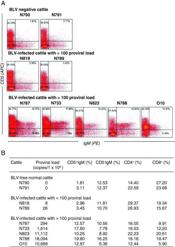
Results: Phenotypic characterization of five BLV-infected but clinically normal cattle with a proviral load of > 100 copies per 1 × 10⁵ cells identified a high percentage of CD5+ IgM+ cells (but not CD5- IgM+ B cells, CD4+ T cells, or CD8+T cells). These lymphocyte subpopulations were purified from three out of five cattle by cell sorting or using magnetic beads, and the BLV proviral load was estimated using BLV-CoCoMo-qPCR. The CD5+ IgM+ B cell population in all animals harbored a higher BLV proviral load than the other cell populations. The copy number of proviruses infecting CD5- IgM+ B cells, CD4+ cells, and CD8+ T cells (per 1 ml of blood) was 1/34 to 1/4, 1/22 to 1/3, and 1/31 to 1/3, respectively, compared with that in CD5+ IgM+ B cells. Moreover, the BLV provirus remained integrated into the genomic DNA of CD5+ IgM+ B cells, CD5- IgM+ B cells, CD4+ T cells, and CD8+ T cells, even in BLV-infected cattle with a proviral load of <100 copies per 105 cells.
Conclusions: The results of the recent study showed that, although CD5+ IgM+ B cells were the main cell type targeted in BLV-infected but clinically normal cattle, CD5- IgM+ B cells, CD4+ cells, and CD8+ T cells were infected to a greater extent than previously thought.
Figures
Similar articles
-
The prevalence of proviral bovine leukemia virus in peripheral blood mononuclear cells at two subclinical stages of infection.J Virol. 1996 Apr;70(4):2178-83. doi: 10.1128/JVI.70.4.2178-2183.1996. J Virol. 1996. PMID: 8642640 Free PMC article.
-
BLV-CoCoMo-qPCR: a useful tool for evaluating bovine leukemia virus infection status.BMC Vet Res. 2012 Sep 21;8:167. doi: 10.1186/1746-6148-8-167. BMC Vet Res. 2012. PMID: 22995575 Free PMC article.
-
BLV-CoCoMo-qPCR: Quantitation of bovine leukemia virus proviral load using the CoCoMo algorithm.Retrovirology. 2010 Nov 2;7:91. doi: 10.1186/1742-4690-7-91. Retrovirology. 2010. PMID: 21044304 Free PMC article.
-
Detrimental effect of bovine leukemia virus (BLV) on the immunological state of cattle.Vet Immunol Immunopathol. 1996 Nov;54(1-4):293-302. doi: 10.1016/s0165-2427(96)05706-6. Vet Immunol Immunopathol. 1996. PMID: 8988875 Review.
-
Viral oncogenesis of δ-retroviruses, HTLV-1 and BLV, and recent advances in its diagnosis.Virology. 2025 Apr;605:110461. doi: 10.1016/j.virol.2025.110461. Epub 2025 Feb 21. Virology. 2025. PMID: 40015031 Review.
Cited by
-
Mapping of CD4+ T-cell epitopes in bovine leukemia virus from five cattle with differential susceptibilities to bovine leukemia virus disease progression.Virol J. 2019 Dec 16;16(1):157. doi: 10.1186/s12985-019-1259-9. Virol J. 2019. PMID: 31842930 Free PMC article.
-
Comprehensive Comparison of Novel Bovine Leukemia Virus (BLV) Integration Sites between B-Cell Lymphoma Lines BLSC-KU1 and BLSC-KU17 Using the Viral DNA Capture High-Throughput Sequencing Method.Viruses. 2022 May 7;14(5):995. doi: 10.3390/v14050995. Viruses. 2022. PMID: 35632737 Free PMC article.
-
Importance of continuous monitoring of bovine leukaemia virus infection on farms in bovine leukaemia virus-endemic areas of Japan.Vet Rec Open. 2025 Jun 27;12(2):e70014. doi: 10.1002/vro2.70014. eCollection 2025 Dec. Vet Rec Open. 2025. PMID: 40585308 Free PMC article.
-
BoLA-DRB3 Polymorphism Controls Proviral Load and Infectivity of Bovine Leukemia Virus (BLV) in Milk.Pathogens. 2022 Feb 5;11(2):210. doi: 10.3390/pathogens11020210. Pathogens. 2022. PMID: 35215153 Free PMC article.
-
BoLA-DRB3 Polymorphism is Associated with Differential Susceptibility to Bovine Leukemia Virus-Induced Lymphoma and Proviral Load.Viruses. 2020 Mar 22;12(3):352. doi: 10.3390/v12030352. Viruses. 2020. PMID: 32235771 Free PMC article.
References
-
- Aida Y, Miyasaka M, Okada K, Onuma M, Kogure S, Suzuki M, Minoprio P, Levy D, Ikawa Y. Further phenotypic characterization of target cells for bovine leukemia virus experimental infection in sheep. Am J Vet Res. 1989;50(11):1946–1951. - PubMed
-
- Djilali S, Parodi AL, Levy D, Cockerell GL. Development of leukemia and lymphosarcoma induced by bovine leukemia virus in sheep: a hematopathological study. Leukemia. 1987;1(11):777–781. - PubMed
-
- Stott ML, Thurmond MC, Dunn SJ, Osburn BI, Stott JL. Integrated bovine leukosis proviral DNA in T helper and T cytotoxic/suppressor lymphocytes. J Gen Virol. 1991;72(Pt 2):307–315. - PubMed
Publication types
MeSH terms
Substances
LinkOut - more resources
Full Text Sources
Other Literature Sources
Research Materials
