. 2013 Jul 1;10(7):2642-52.
doi: 10.1021/mp400075f. Epub 2013 May 29.

N-terminal truncation of an isolated human IgG1 CH2 domain significantly increases its stability and aggregation resistance

Affiliations

N-terminal truncation of an isolated human IgG1 CH2 domain significantly increases its stability and aggregation resistance

Rui Gong et al. Mol Pharm. .

Abstract

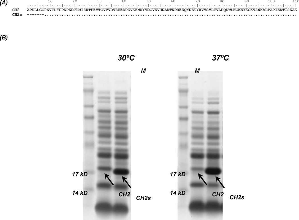
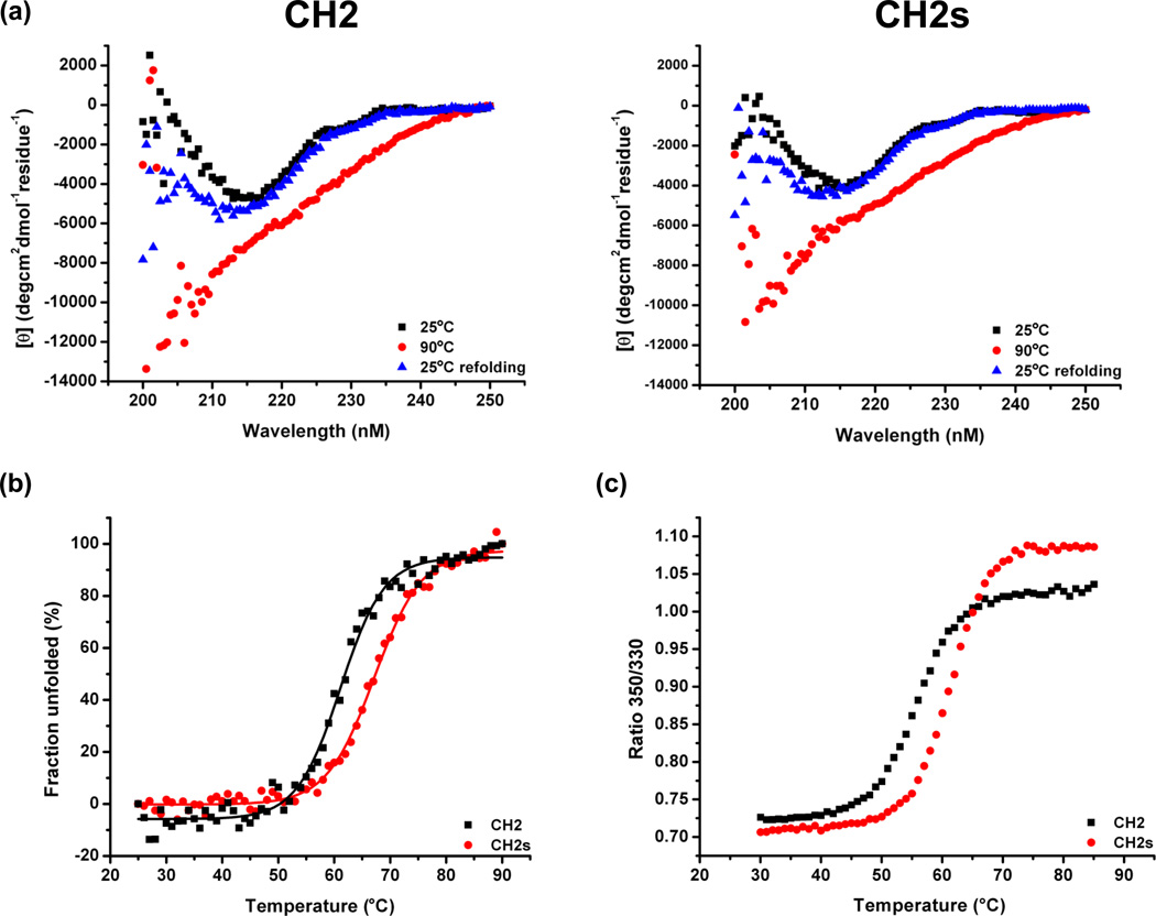
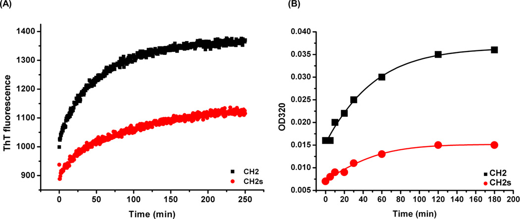
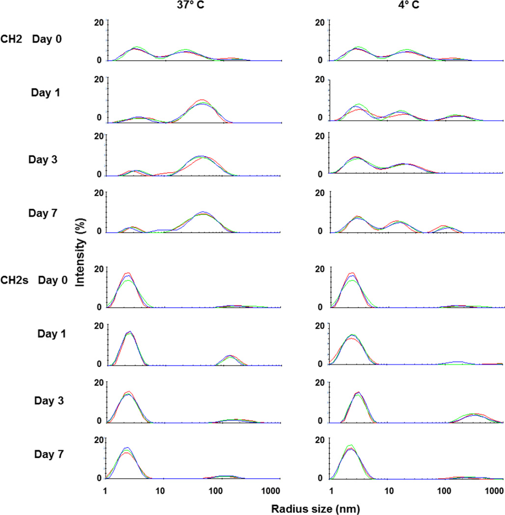
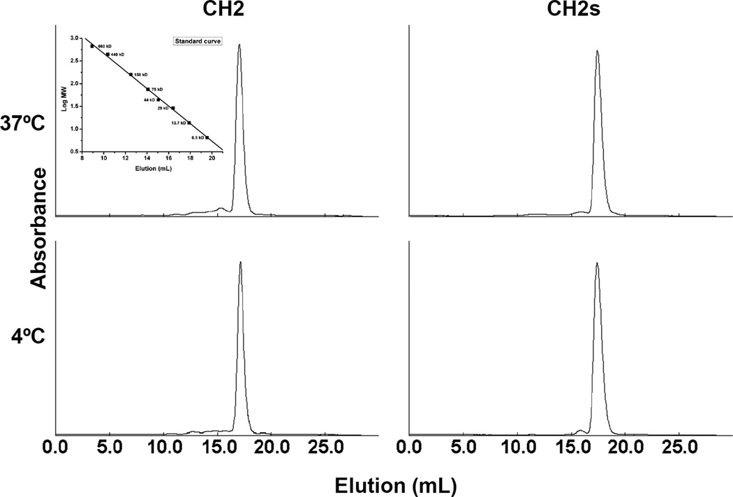
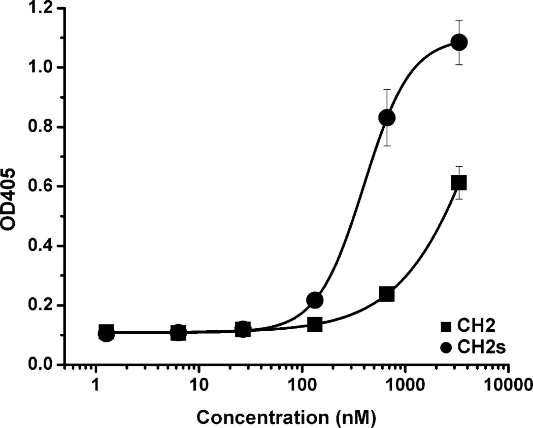
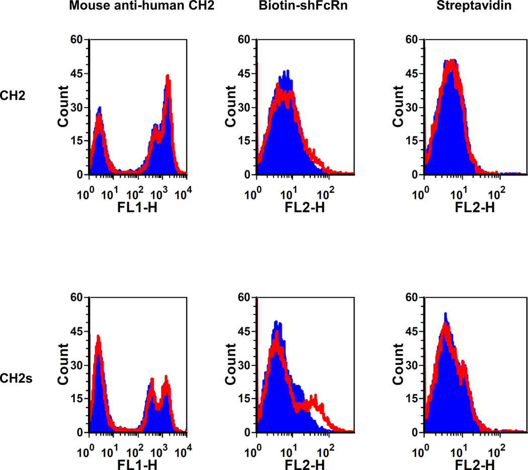
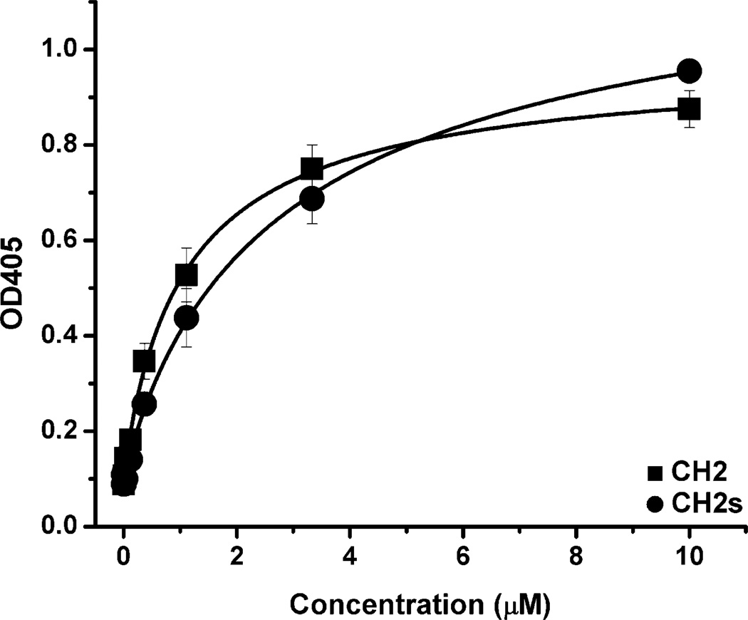
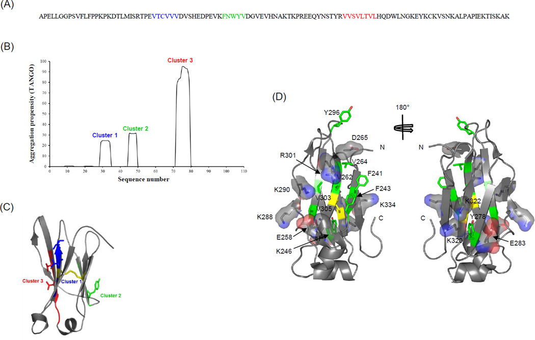
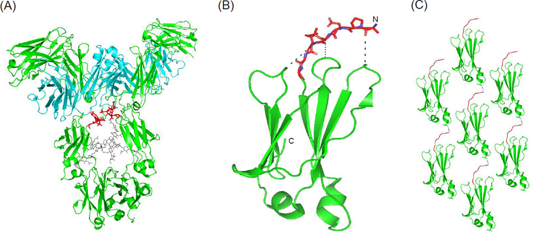
Isolated human immunoglobulin G (IgG) CH2 domains are promising scaffolds for novel candidate therapeutics. Unlike other human IgG domains, CH2 is not involved in strong interchain interactions, and isolated CH2 is relatively stable. However, isolated single CH2 is prone to aggregation. In native IgG and Fc molecules, the N-terminal residues of CH2 from the two heavy chains interact with each other and form hinge regions. By contrast, the N-terminal residues are highly disordered in isolated CH2. We have hypothesized that the removal of the CH2 N-terminal residues may not only increase its stability but also its aggregation resistance. To test this hypothesis we constructed a shortened variant of IgG1 CH2 (CH2s) where the first seven residues of the N-terminus were deleted. We found that the thermal stability of CH2s was increased by 5 °C compared to CH2. Importantly, we demonstrated that CH2s is significantly less prone to aggregation than CH2 as measured by Thioflavin T (ThT) fluorescence, turbidity, and light scattering. We also found that the CH2s exhibited pH-dependent binding to a soluble single-chain human neonatal Fc receptor (shFcRn) which was significantly stronger than the very weak binding of CH2 to shFcRn as measured by flow cytometry. Computer modeling suggested a possible mode of CH2 aggregation involving its N-terminal residues. Therefore, deletion of the N-terminal residues could increase drugability of CH2-based therapeutic candidates. This strategy to increase stability and aggregation resistance could also be applicable to other Ig-related proteins.


Figures

Figure 1
Figure 1
Design and expression of CH2 and CH2s. (A) Amino acid sequence alignment of wide-type CH2 (NCB Accession No. J00228) with CH2s. (B) Comparison of the soluble expression of CH2 and CH2s at 30°C and 37°C.
Figure 2
Figure 2
Secondary structure and stability of CH2 and CH2s measured by circular dichroism (CD). (A) Folding curve at 25°C (black), unfolding at 90°C (red) and refolding (blue) at 25°C. (B) The fraction folded of the protein (ff) was calculated as ff = ([θ] -[θM])/([θT] -[θM]). [θT] and [θM] were the mean residue ellipticities at 216 nm of folded state at 25°C and unfolded state of 90°C. The Tm values (57.7°C and 62.5°C for CH2 and CH2s respectively) from were determined by the first derivative [d(Fraction folded)/dT] with respect to temperature (T). (C) Temperature induced unfolding of CH2 and CH2s. The fluorescence intensity ratio of 350/330 indicated that tryptophan residues were exposed as the CH2 and CH2s unfolded. The calculated Tm values were 56.2°C and 61.3°C for CH2 and CH2s respectively.
Figure 3
Figure 3
Comparison of aggregation propensity between CH2 and CH2s. (A) ThT binding experiment at room temperature. The rate of increase of fluorescence intensity in CH2 (black) was faster than that in CH2s (red). (B) Turbidity assay of CH2 (black) and CH2s (red) after 50°C incubation at different time points. OD320 of CH2 increased faster than that for CH2s.
Figure 4
Figure 4
Measurement of CH2 and CH2s aggregation after incubation at 37°C and 4°C by dynamic light scattering (DLS). CH2 had two major peaks at day 0 while CH2s had only one peak indicating that the populations of CH2 molecules were not uniform and reflected possible oligomer formation after purification.
Figure 5
Figure 5
Estimation of oligomer formation of CH2 and CH2s after 7-day incubation at 37°C and 4°C by SEC. Only monomeric peak was observed in both CH2 and CH2s indicating that the formation of oligomer might be reversible. The insertion is standard curve.
Figure 6
Figure 6
Binding of CH2 (■) and CH2s (●) to an anti-human CH2 Fab m01m1. The EC50s of m01m1 to CH2 and CH2s were >1400 nM and 392 nM, respectively, indicating conformational differences.
Figure 7
Figure 7
Binding of yeast-expressed CH2 and CH2s to shFcRn at pH6 (red) and pH7.4 (blue). Very slight fluorescence intensity shift occurred in the case of CH2 indicating very weak binding to shFcRn. Stronger fluorescence intensity shift was observed in the case of CH2s indicating enhanced binding to shFcRn. The expression of CH2 and CH2s was tested by the corresponding antibodies. PE-streptavidin was used as negative control.
Figure 8
Figure 8
Binding of CH2 and CH2s to human serum albumin (HSA) measured by ELISA. (■) binding of CH2 to HSA (EC50 ≈ 0.9 µM); (●) binding of CH2s to HSA (EC50 ≈ 1.7 µM).
Figure 9
Figure 9
Prediction of aggregation prone regions (APRs) in CH2. (A) Three major APRs, cluster 1–3, are mapped on the CH2 amino acid sequence and highlighted by blue, green and red, respectively, as predicted by TANGO method. (B) The TANGO profile for CH2 showing the sequence number on X-axis and aggregation propensity (%) on Y-axis. (C) Mapping of APRs, clusters 1–3, on the three-dimensional structure of CH2 domain. (D) Ribbon diagrams of CH2 structure illustrating the distribution of charged residues as sticks embedded in translucent surfaces, and hydrophobic residues in green sticks with atom colors are shown. The location of disulfide bond is colored in yellow.
Figure 10
Figure 10
(A) An intact IgG crystal structure is shown for highlighting the N-terminal residues of CH2 domains, colored in red sticks (PDB code 1HZH). The carbohydrates moieties are shown in gray sticks. (B) A ribbon cartoon diagram of the CH2 domain with modeled N-terminal residues (in orange sticks and colored by atoms) showing the proximity of the N-terminal residues to the loop regions of CH2. (C) A crystal packing view of CH2 monomers in the lattice indicates intermolecular contacts between the N-terminal residues and helical region of different CH2 monomers as shown by dots. Surface-exposed hydrophobic residue Leu253 at a helical region involved in the intermolecular interaction is shown as sticks.

References

    1. Waldmann TA. Immunotherapy: past, present and future. Nat Med. 2003;9(3):269–277. - PubMed
    1. Casadevall A, Dadachova E, Pirofski LA. Passive antibody therapy for infectious diseases. Nature reviews Microbiology. 2004;2(9):695–703. - PubMed
    1. Schrama D, Reisfeld RA, Becker JC. Antibody targeted drugs as cancer therapeutics. Nature reviews Drug discovery. 2006;5(2):147–159. - PubMed
    1. Carter PJ. Potent antibody therapeutics by design. Nat Rev Immunol. 2006;6(5):343–357. - PubMed
    1. Dimitrov DS, Marks JD. Therapeutic antibodies: current state and future trends--is a paradigm change coming soon? Methods Mol Biol. 2009;525(1–27):xiii. - PMC - PubMed

Publication types