

Optimization of human corneal endothelial cell culture: density dependency of successful cultures in vitro
- PMID: 23641909
- PMCID: PMC3659058
- DOI: 10.1186/1756-0500-6-176
Optimization of human corneal endothelial cell culture: density dependency of successful cultures in vitro
Abstract
Background: Global shortage of donor corneas greatly restricts the numbers of corneal transplantations performed yearly. Limited ex vivo expansion of primary human corneal endothelial cells is possible, and a considerable clinical interest exists for development of tissue-engineered constructs using cultivated corneal endothelial cells. The objective of this study was to investigate the density-dependent growth of human corneal endothelial cells isolated from paired donor corneas and to elucidate an optimal seeding density for their extended expansion in vitro whilst maintaining their unique cellular morphology.
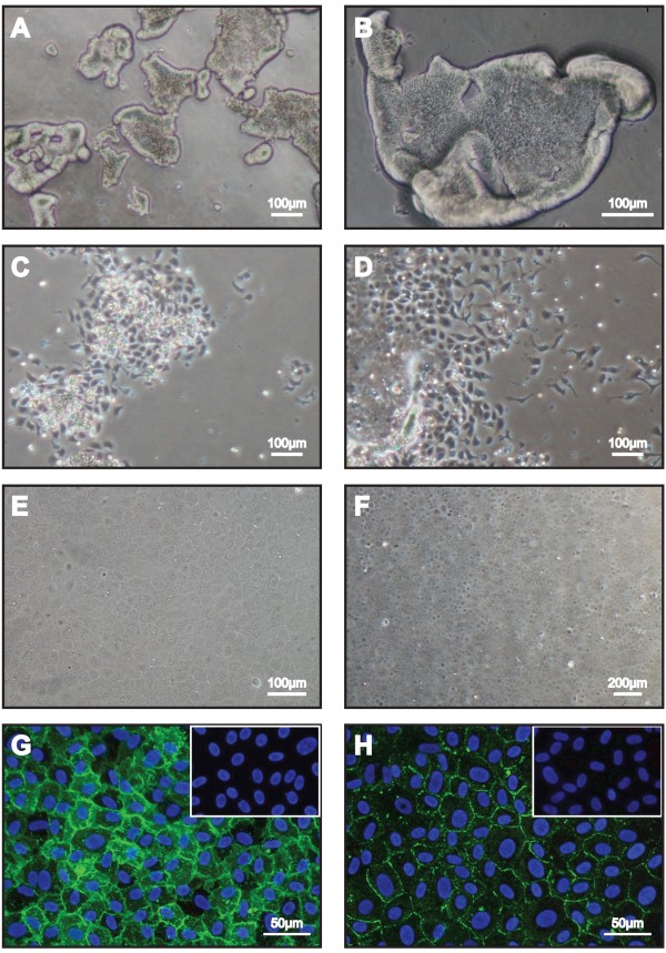
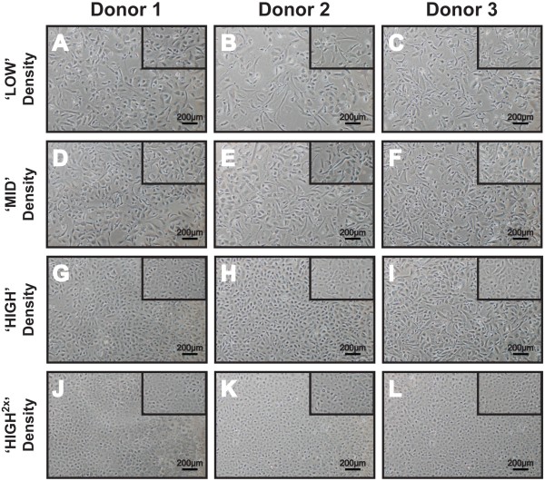
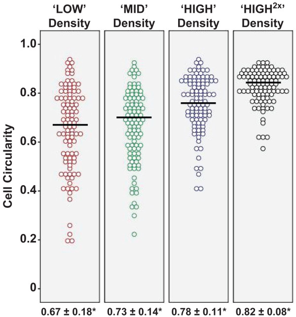
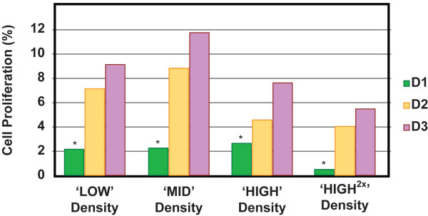
Results: Established primary human corneal endothelial cells were propagated to the second passage (P2) before they were utilized for this study. Confluent P2 cells were dissociated and seeded at four seeding densities: 2,500 cells per cm2 ('LOW'); 5,000 cells per cm2 ('MID'); 10,000 cells per cm2 ('HIGH'); and 20,000 cells per cm2 ('HIGH(×2)'), and subsequently analyzed for their propensity to proliferate. They were also subjected to morphometric analyses comparing cell sizes, coefficient of variance, as well as cell circularity when each culture became confluent. At the two lower densities, proliferation rates were higher than cells seeded at higher densities, though not statistically significant. However, corneal endothelial cells seeded at lower densities were significantly larger in size, heterogeneous in shape and less circular (fibroblastic-like), and remained hypertrophic after one month in culture. Comparatively, cells seeded at higher densities were significantly homogeneous, compact and circular at confluence. Potentially, at an optimal seeding density of 10,000 cells per cm2, it is possible to obtain between 10 million to 25 million cells at the third passage. More importantly, these expanded human corneal endothelial cells retained their unique cellular morphology.
Conclusions: Our results demonstrated a density dependency in the culture of primary human corneal endothelial cells. Sub-optimal seeding density results in a decrease in cell saturation density, as well as a loss in their proliferative potential. As such, we propose a seeding density of not less than 10,000 cells per cm2 for regular passage of primary human corneal endothelial cells.
Figures
References
-
- Klyce SD, Beuerman RW. In: The Cornea. Kaufman HE, Barron BA, McDonald MB, Waltman SR, editor. New York: Churchill Livingstone; 1988. Structure and function of the cornea; pp. 3–28.
-
- Geroski DH, Matsuda M, Yee RW, Edelhauser HF. Pump function of the human corneal endothelium. Effects of age and cornea guttata. Ophthalmology. 1985;92(6):759–763. - PubMed
Publication types
MeSH terms
LinkOut - more resources
Full Text Sources
Other Literature Sources
Research Materials
