. 2013 May 4:8:9.
doi: 10.1186/1749-8104-8-9.

Development and specification of cerebellar stem and progenitor cells in zebrafish: from embryo to adult

Affiliations

Development and specification of cerebellar stem and progenitor cells in zebrafish: from embryo to adult

Jan Kaslin et al. Neural Dev. .

Abstract

Background: Teleost fish display widespread post-embryonic neurogenesis originating from many different proliferative niches that are distributed along the brain axis. During the development of the central nervous system (CNS) different cell types are produced in a strict temporal order from increasingly committed progenitors. However, it is not known whether diverse neural stem and progenitor cell types with restricted potential or stem cells with broad potential are maintained in the teleost fish brain.

Results: To study the diversity and output of neural stem and progenitor cell populations in the zebrafish brain the cerebellum was used as a model brain region, because of its well-known architecture and development. Transgenic zebrafish lines, in vivo imaging and molecular markers were used to follow and quantify how the proliferative activity and output of cerebellar progenitor populations progress. This analysis revealed that the proliferative activity and progenitor marker expression declines in juvenile zebrafish before they reach sexual maturity. Furthermore, this correlated with the diminished repertoire of cell types produced in the adult. The stem and progenitor cells derived from the upper rhombic lip were maintained into adulthood and they actively produced granule cells. Ventricular zone derived progenitor cells were largely quiescent in the adult cerebellum and produced a very limited number of glia and inhibitory inter-neurons. No Purkinje or Eurydendroid cells were produced in fish older than 3 months. This suggests that cerebellar cell types are produced in a strict temporal order from distinct pools of increasingly committed stem and progenitor cells.

Conclusions: Our results in the zebrafish cerebellum show that neural stem and progenitor cell types are specified and they produce distinct cell lineages and sub-types of brain cells. We propose that only specific subtypes of brain cells are continuously produced throughout life in the teleost fish brain. This implies that the post-embryonic neurogenesis in fish is linked to the production of particular neurons involved in specific brain functions, rather than to general, indeterminate growth of the CNS and all of its cell types.


Figures

Figure 1
Figure 1
Overview of the cerebellar architecture in zebrafish. (A) In the adult zebrafish brain neural stem cells are abundant and distributed in distinct topological clusters along the whole rostro-caudal brain axis; (B) A schematic coronal section showing the anatomy of the zebrafish cerebellum. The cerebellum has a simple laminar three layered architecture consisting of a molecular layer (ML), Purkinje cell layer (PL) and a granule cell layer (GL). The granule layer consists of excitatory granule cells and inhibitory Golgi neurons. The Purkinje cell layer contains Purkinje neurons (PN), Bergmann glia (G), and excitatory eurydendroid cells (E). The ML mainly consists of nerve fibers and scattered inhibitory stellate cells. Boxed region. Neural progenitors are maintained in the dorsomedial part of the cerebellum around a remnant of the IVth ventricle (the cerebellar recessus, CR). The progenitors give rise to granule neurons in a distinct outside-in fashion. (1) Polarized neuroepithelial-like progenitors (green) are restricted to the midline of the dorsal cerebellum. The progenitors give rise to rapidly migrating granule precursors (dark green) that initially migrate dorsolaterally. After reaching the meninge the granule precursors migrate in ventrolateral direction to the GL. (2) Radial glia (light blue) are found close to the midline and they are used as scaffolds during the initial dorsal migration of granule precursors. C) Schematic view of the cerebellar progenitor domains during development. The cerebellar cell types are generated from two germinal zones: the ventricular zone (VZ, orange) and the upper rhombic lip (URL, green). The excitatory neurons are generated from the URL, while inhibitory neurons and glia are generated from the VZ. The cerebellar cell types are produced in a strict temporal order from increasingly committed progenitors. Purkinje neurons and deep cerebellar nuclei neurons are produced early during development, while inhibitory interneurons, granule cells and glia are produced late.
Figure 2
Figure 2
Distinct cerebellar progenitor populations are established early in the embryo. (A) Expression of ptf1a, DsRed (mRNA) and DsRed (protein) in developing and juvenile Ptf1a:DsRed transgenic fish. Ptf1a, DsRed and DsRed show similar expression patterns; (B) In vivo expression of DsRed and Egfp in the embryonic and juvenile Ptf1a:DsRed and Olig2:egfp transgenic fish. Overlapping Egfp and DsRed expression is seen in the VZ of the cerebellar primordium; (C) Nestin:egfp+ (green) and Ptf1a:DsRed+ (red) progenitors in the cerebellar primordium. The Nestin:egfp labels cells in the URL, while Ptf1a:DsRed line labels cells in the VZ. Two days post-fertilization Nestin:egfp+ and Ptf1a:DsRed+ cells form distinct populations in the URL and VZ of the cerebellar primordium; (D) A dorsal overview of Nestin:egfp+ (green) and Ptf1a:DsRed+ (red) progenitors in the hindbrain of a 5-day-old larval zebrafish. Two distinct progenitor domains are visible in the cerebellum (the junction is indicated with an arrow). Ptf1a:DsRed+ cells localize along the ventricular zone of the IVth ventricle (the VZ domain is labeled with a hatched orange line), while Nestin:egfp+ cells localize in the URL (hatched green line). MHB: Mid-hindbrain boundary; LRL: Lower rhombic lip.
Figure 3
Figure 3
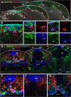
Proliferative activity of cerebellar progenitors diminishes during juvenile stages. (A) Confocal maximum projection of cerebellar cross sections at a similar level of juvenile, young and adult cerebellum. Proliferating cells are labeled with proliferating cell nuclear antigen (PCNA) in blue. Nestin:egfp+ cells are green, Ptf1a:DsRed+ cells are red and the general tissue morphology is depicted with DAPI staining in grey. Yellow arrows depict the distinct niche in the URL where proliferating Nestin:egfp+/PCNA+ progenitors are located. White arrows depict Ptf1a:DsRed+ cells that localize to the ventral part of the IVth ventricle and to the cerebellar parenchyma. The majority of the parenchymal Ptf1a:DsRed+ cells are PCNA- while most of the Ptf1a:DsRed+ cells at the VZ close to the ventricle are PCNA+. (B-C) High magnification single confocal planes of the boxed areas in A. (B) A proliferating Ptf1a:DsRed+ /PCNA+ progenitor at the IVth ventricle (white arrow) and many proliferating Nestin:egfp+/PCNA+ cells in the URL (yellow arrow); (C) A proliferating Ptf1a:DsRed+ cell in the cerebellar parenchyma (white arrow); (D) Confocal maximum projections of juvenile and adult cerebellar cross sections showing a decline in VZ progenitor activity. (E) High magnification of boxed area in D. Proliferating Ptf1a:DsRed+ (red) and Olig2:egfp+ (green) cells are found in VZ and the cerebellar parenchyma in a two-week-old juvenile fish. Note the absence of Ptf1a:DsRed+ and Olig2:egfp+ cells among the PCNA+ cells in the URL. The white arrow depicts the Ptf1a:DsRed+ and Olig2:egfp+cells laminating from the ventricular surface into the parenchyma; (F) Ptf1a:DsRed+ cells adjacent to proliferating PCNA+ cells in a one-month-old fish; (G) Proliferating Ptf1a:DsRed+ and Olig2:egfp+ in the VZ of the fourth ventricle (arrow) in a one-month-old fish. The white arrow shows the Ptf1a:DsRed+ and Olig2:egfp+ cells laminating from the ventricular surface into the parenchyma.
Figure 4
Figure 4
Quantification of the proliferative activity of cerebellar progenitors. The number of proliferating Nestin:egfp+ progenitors are significantly reduced during juvenile stages but notable progenitor activity is still detected in the adult and aging brain (P <0.001, n = 5). A significant loss of the proliferative activity of Ptf1a:DsRed+ and Olig2:egfp+ cells takes place during juvenile stage and the activity is almost exhausted in the adult (Ptf1a:DsRed: P <0.001, n = 7; Olig2:egfp: P <0.001, n = 5). The proliferating Olig2:egfp cells show a similar decline in the proliferative activity at the ventricle and in the brain parenchyma as the Ptf1a:DsRed+ cells.
Figure 5
Figure 5
Cerebellar progenitors gives rise to distinct cell lineages. (A) Co-localization (arrow) of Ptf1a:DsRed (blue) and GABA, PV or ZII (green) in inhibitory neurons in the cerebellum of a 5-day-old larvae; (B,C) Ptf1a:DsRed+ cells showing morphologies of differentiating stellate and Golgi neurons; (D) A PV- (red) Ptf1a:DsRed+ (green) and HU C/D+ (blue) stellate cell in the ML; (E) Co-localization (arrows) of Ptf1a:DsRed (blue) and GABA (red) cells in the cerebellum of a 1-month-old juvenile zebrafish; (F) Cross section of the juvenile cerebellum showing abundant co-localization (white arrow) of Ptf1a:DsRed (red) and Pax2 (green); (G) Overview of a cross section of the adult cerebellum showing Pax2 (green) labeled Golgi neurons in the GL and PL. Purkinje neurons in the PL are labeled with parvalbumin (red); (H) Co-localization (arrow) of Olig2:egfp (green) and HU C/D (blue) cell in the PL of an adult zebrafish.
Figure 6
Figure 6
Morphology of Ptf1a:DsRed cells in the VZ. (A) High magnification of a ventricularly located Ptf1a:DsRed+ cell with radial morphology in a juvenile zebrafish; (B) A Ptf1a:DsRed+ cell (red) with a radial morphology located at the ventricle, ventral to the Nestin:egfp+ cells (green) in the URL. Ventricle and apical junctions are outlined with ZO-1 (blue); (C) Ptf1a:DsRed+ and gfap:egfp+ cells (green) at the ventricle of the VZ. Proliferating cells in the progenitor niche are labeled with PCNA (blue) and glia are labeled by gfap:egfp (green). The white arrow depicts a proliferating Ptf1a:DsRed+and PCNA+ cell located lateral to the PCNA+ cells in the URL. The yellow arrow depict a horizontally oriented Ptf1a:DsRed+ and gfap:egfp+ cell at the ventricle; (D) Flat horizontally oriented s100β+ cells (red) are lining the VZ of the progenitor niche in adult zebrafish. The ventricle is outlined with the junctional marker ZO-1 (blue); (E) A Ptf1a:DsRed+ Bergmann glia in the adult cerebellum; (F) A Ptf1a:DsRed+ and Nestin:egfp+ Bergmann glia lateral to the progenitor niche (arrow); (G) Co-localization (arrows) of Nestin:egfp (blue) and NeuroD1 (green) in the cerebellum of a 5 day old larvae; (H) No overlap between Nestin:egfp (green, white arrow) cells and PV+ cells (blue, yellow arrow) is seen in the cerebellum of a 5-day-old larvae; (I) Overlapping expression of Atoh1c and Nestin:egfp in the URL. C: Cerebellum, GL: Granule cell layer, URL: Upper rhombic lip, VZ: Ventricular zone.
Figure 7
Figure 7
Generation of cerebellar cell types over time. (A) Quantification of parvalbumin immunopositive cells in the cerebellum of 7- and 14-day-old juvenile zebrafish shows a significant increase of PV+ cells in the cerebellum between 7- and 14-day-old zebrafish (P = 0.0003, n = 4). The pictures show a top view of the cerebellum and PV + cells; (B) An overview of the BrdU pulse chase experiments. To maximize the labeling of dividing cells the zebrafish were given five consecutive 14 hour pulses of BrdU. The fish were euthanized four weeks after the last BrdU pulse; (C) Confocal maximum projections of juvenile and adult cerebellar cross sections showing a notable production of cells in the juvenile and adult zebrafish cerebellum. PV+ Purkinje cells (red) and their processes are seen in in the PL. Scattered s100β+ glia (blue) are seen in the PL and ML. In the juvenile fish high numbers of BrdU+ cells (green) are found both in the GL (orange arrows) and PL (white and yellow arrows). In the adult cerebellum BrdU+ cells are confined to the GL (orange arrows); (D) High magnification of a single confocal plane of the boxed area in C showing a s100β+ glia co-localizing with BrdU (white arrow) and a PV+ Purkinje neuron co-localizing with BrdU (yellow arrow) in a juvenile zebrafish; (E) High magnification maximum projection of the boxed area in C showing s100β+ Bergmann glia lateral to the URL progenitor niche (yellow hatched line) co-localizing with BrdU (white arrows); (F) High magnification of BrdU+ and Pax6+ granule cells (white arrows) in the GL.
Figure 8
Figure 8
Generation of different cerebellar cell types. (A) Confocal maximum projection of a cerebellar cross section in a juvenile zebrafish showing BrdU labeled GABA+ inhibitory neurons (white arrows) and BrdU+/GABA+/PV+ neurons (yellow arrows); (B) High magnification of a single confocal plane showing a neuron co-localizing with BrdU and GABA (white arrow); (C) High magnification of a single confocal plane showing a neuron co-localizing with BrdU, GABA and PV (white arrow); (D) Confocal maximum projection of a cerebellar cross section showing BrdU labeled ZII+ Purkinje neurons (white arrows) and a Pax2+ Golgi neuron (yellow arrow); (E) High magnification of a single confocal plane showing a Golgi neuron co-localizing with BrdU and Pax2 (white arrow); (F) Magnified single confocal plane showing Purkinje neurons co-localizing with BrdU and ZII (white arrows); (G) A rare proliferating oligodendrocyte progenitor (PCNA/Olig2:egfp+) detected in the cerebellar parenchyma (arrow); (H) A putative oligodendrocyte labeled with BrdU+and Olig2:egfp+ detected in the brain parenchyma six weeks after the BrdU pulse; (I) Quantifications of the cell types produced in the cerebellum of juvenile and adult zebrafish after four weeks BrdU pulse chasing. A significant decline of cerebellar inhibitory neuron and glia production between juvenile and adult stages is detected while granule cell production declines during juvenile stages but is still maintained at a high level in the adult zebrafish (n = 5, P <0.001).
Figure 9
Figure 9
Summary of the cerebellar progenitors and the progenitor niche in zebrafish. (A) During larval and juvenile stages the URL and a portion of the VZ together with a part of IVth ventricle (cerebellar recessus, CR) is displaced from the rest of the ventricle through tissue growth (red opposing arrows). The neural progenitors in the URL (dorsal part) remain active throughout life, while the VZ progenitors (ventral part) largely become quiescent in the adult. During larval and juvenile stages progenitors delaminate from the VZ into parenchyma. The highest density of proliferating VZ progenitors is found at the interface of the caudal lobe (best seen in C); (B) Schematic view of the cellular arrangement of the cerebellar progenitor niche. 1. Granule cell stem and progenitor cells (green) are found dorsal to the cerebellar recessus in the URL. The granule precursors migrate to the GL and differentiate into granule neurons. 2. Ventricular zone derived progenitors (orange) are found ventral to the recessus. Proliferating and differentiating VZ progenitors migrate to the cerebellar parenchyma during larval and juvenile stages. Most of the cells in VZ of the mature cerebellum express glial and ependymal markers and display epithelial-like morphology. Bergmann glia and inhibitory inter-neurons are produced at the interface between the URL and VZ. 3. Very rare progenitors reside in the cerebellar parenchyma. (C) Schematic parasagittal view of the cerebellum and the distribution of progenitors; (D) The URL derived progenitors remain active into adulthood, while the proliferative activity of VZ derived progenitors virtually is exhausted during juvenile stages. Purkinje neurons and Eurydendroid cells are produced up to 1 month. In sexually mature zebrafish only few inhibitory neurons and glia are produced. Production of granule neurons is maintained throughout life in zebrafish.

Similar articles

Cited by

References

    1. Merkle FT, Mirzadeh Z, Alvarez-Buylla A. Mosaic organization of neural stem cells in the adult brain. Science. 2007;317:381–384. - PubMed
    1. Kelsch W, Mosley CP, Lin CW, Lois C. Distinct mammalian precursors are committed to generate neurons with defined dendritic projection patterns. PLoS Biol. 2007;5:e300. - PMC - PubMed
    1. Hack MA, Saghatelyan A, de Chevigny A, Pfeifer A, Ashery-Padan R, Lledo PM, Gotz M. Neuronal fate determinants of adult olfactory bulb neurogenesis. Nat Neurosci. 2005;8:865–872. - PubMed
    1. Shen Q, Wang Y, Dimos JT, Fasano CA, Phoenix TN, Lemischka IR, Ivanova NB, Stifani S, Morrisey EE, Temple S. The timing of cortical neurogenesis is encoded within lineages of individual progenitor cells. Nat Neurosci. 2006;9:743–751. - PubMed
    1. Encinas JM, Michurina TV, Peunova N, Park JH, Tordo J, Peterson DA, Fishell G, Koulakov A, Enikolopov G. Division-coupled astrocytic differentiation and age-related depletion of neural stem cells in the adult hippocampus. Cell Stem Cell. 2011;8:566–579. - PMC - PubMed

Publication types

LinkOut - more resources