. 2013 May 4:10:58.
doi: 10.1186/1742-2094-10-58.

Microglial activation decreases retention of the protease inhibitor saquinavir: implications for HIV treatment

Affiliations

Microglial activation decreases retention of the protease inhibitor saquinavir: implications for HIV treatment

Shannon Dallas et al. J Neuroinflammation. .

Abstract

Background: Active HIV infection within the central nervous system (CNS) is confined primarily to microglia. The glial cell compartment acts as a viral reservoir behind the blood-brain barrier. It provides an additional roadblock to effective pharmacological treatment via expression of multiple drug efflux transporters, including P-glycoprotein. HIV/AIDS patients frequently suffer bacterial and viral co-infections, leading to deregulation of glial cell function and release of pro-inflammatory mediators including cytokines, chemokines, and nitric oxide.

Methods: To better define the role of inflammation in decreased HIV drug accumulation into CNS targets, accumulation of the antiretroviral saquinavir was examined in purified cultures of rodent microglia exposed to the prototypical inflammatory mediator lipopolysaccharide (LPS).

Results: [(3)H]-Saquinavir accumulation by microglia was rapid, and was increased up to two-fold in the presence of the specific P-glycoprotein inhibitor, PSC833. After six or 24 hours of exposure to 10 ng/ml LPS, saquinavir accumulation was decreased by up to 45%. LPS did not directly inhibit saquinavir transport, and did not affect P-glycoprotein protein expression. LPS exposure did not alter RNA and/or protein expression of other transporters including multidrug resistance-associated protein 1 and several solute carrier uptake transporters.

Conclusions: The decrease in saquinavir accumulation in microglia following treatment with LPS is likely multi-factorial, since drug accumulation was attenuated by inhibitors of NF-κβ and the MEK1/2 pathway in the microglia cell line HAPI, and in primary microglia cultures from toll-like receptor 4 deficient mice. These data provide new pharmacological insights into why microglia act as a difficult-to-treat viral sanctuary site.


Figures

Figure 1
Figure 1
Accumulation of saquinavir by HAPI microglia in the presence of the P-glycoprotein inhibitor PSC833. (A) [3H]saquinavir (50 nM) accumulation by HAPI cells was measured in the presence (closed circles) or absence (open circles) of PSC833 (5 μM) at 0.5, 1, 2, 5, 10, 30, 60 and 120 minutes, at 37°C. (B) [3H]saquinavir (50 nM) accumulation by HAPI microglia increased significantly, in a dose dependent manner, in the presence of excess unlabelled saquinavir (1 to 10 μM). Each data point represents mean ± SE of at least three separate experiments with three replicates included per experiment; *P < 0.05, significantly different from control.
Figure 2
Figure 2
Effect of LPS on [3H]saquinavir accumulation and inflammatory mediator release by HAPI microglia cells. (A) Following 24 hours of incubation at increasing LPS concentrations (1 to 10 ng/ml) (black bars), [3H]saquinavir (50 nM) accumulation in HAPI cells was determined at one hour (37°C) using scintillation counting. The accumulation was dose-dependent at concentrations higher than 2.5 ng/ml LPS. (B) Accumulation of [3H]saquinavir (50 nM) for one hour following addition of LPS into the transport buffer. Each data point represents mean ± SE of at least three separate experiments with three replicates included per experiment; *P < 0.05, significantly different from control.
Figure 3
Figure 3
Interactions with P-glycoprotein in HAPI microglia only partially explain the LPS-mediated changes in saquinavir accumulation. (A) Following 24 hours LPS incubation (10 ng/ml), accumulation of [3H]saquinavir (50 μM) at one hour (37°C) was measured in the presence and absence of the P-glycoprotein inhibitor PSC833 (5 μM). (B) By subtracting the LPS-untreated values (control and PSC833 inhibited; white bars) from the LPS-treated cells (LPS control and PSC833 inhibited; black bars) the P-glycoprotein specific/sensitive component of transport was determined. Each data point represents mean ± SE of at least three separate experiments with three replicates included per experiment; *P < 0.05, significantly different from LPS-untreated control cells.
Figure 4
Figure 4
Protein expression of P-glycoprotein and Mrp1 in HAPI cells following 6 or 24 hour LPS incubation. (A) HAPI microglia were incubated for 6 or 24 hours in the presence of increasing concentrations of LPS (1 to 10 ng/ml). At the end of the incubation period, crude membrane fractions from the cell lysates or from rat kidney brush border membranes (positive P-glycoprotein control) were prepared as indicated in the text, and immunoblotting of P-glycoprotein was performed. The proteins (1 or 40 μg each) were separated on NuPage 7% sodium-acetate gels, transferred to polyvinylidene difluoride membranes electrophoretically and incubated with a P-glycoprotein monoclonal antibody, C219 (1:100), followed by an anti-mouse HRPO-secondary (1:1,000). (B) Immunoblotting of Mrp1 in HAPI microglia treated for 6 or 24 hours with increasing concentrations of LPS (1 to 10 ng/ml) was undertaken in a similar manner to P-glycoprotein except that the proteins (10 μg) were separated on NuPage 4 to 12% Bis-Tris gels, and incubated with a Mrp1 monoclonal antibody, MRPr1 (1:100), followed by an anti-rat HRPO-secondary (1:1,000). Whole rat kidney lysate (10 μg) was used as a positive Mrp1 control. Representative immunoblots are shown.
Figure 5
Figure 5
Decreased saquinavir accumulation by HAPI microglia in the presence of LPS is attenuated in TLR4 deficient mice. (A) Primary microglia were isolated from TLR wild-type (TLR4 WT) and TLR4 deficient (TLR4−/−) mice, as described. In primary microglia isolated from TLR4 WT mice, incubation with 10 ng/ml LPS for 24 hours decreased saquinavir significantly, that is, by approximately 16%. This effect was attenuated in microglia derived from the TLR4−/− deficient mice, where there was no difference between LPS-treated and untreated deficient mice or the untreated WT mice. *P < 0.05, significantly different from untreated TLR WT controls. (B) Primary microglia derived from Wistar, and (C) Fisher rats incubated with 10 ng/ml LPS for 24 hours at 37°C also showed a significant decrease in saquinavir accumulation at one hour. *P < 0.05, significantly different from untreated control cells.
Figure 6
Figure 6
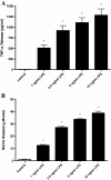
TNF-α and nitrite release by HAPI cells following LPS exposure. Release of TNF-α (A) and nitrite (B) by HAPI cells ino the medium was measured following 24 hours LPS incubation at 1 to 10 ng/ml. Each data point represents mean ± SE of at least three separate experiments with three replicates included per experiment; *P < 0.05, significantly different from untreated control (open bars).
Figure 7
Figure 7
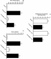
Saquinavir accumulation and inflammatory mediator release following incubation of HAPI cells with a cell permeable NF-κβ inhibitor. (A) Accumulation of [3H]saquinavir (50 nM) at one hour (37°C) following 24 hours incubation with 10 ng/ml LPS and the NF-κβ inhibitor SN50 (10 μM) or inactive control peptide SN50I (10 μM). Release of TNF-α (B) and nitrite (C) into the medium following 24 hours incubation with 10 ng/ml LPS in the presence of the NF-κβ inhibitor SN50 (10 μM) or control peptide SN50I (10 μM) was also measured under the same experimental conditions. Each data point represents mean ± SE of at least three separate experiments with three replicates included per experiment; *P < 0.05, significantly different from untreated control cells; #P < 0.05, significantly different from 10 ng/ml LPS and 10 μM inactive SN50I + LPS.
Figure 8
Figure 8
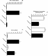
Effect of MEK1/2 inhibitor on saquinavir accumulation, TNF-α and nitrite release. Saquinavir accumulation and inflammatory mediator release was determined following incubation of HAPI cells with a cell permeable MEK1/2 inhibitor U0126 (A). Accumulation of [3H]saquinavir (50 nM) in the presence and absence of 10 ng/ml LPS and 10 μM U0126 at one hour at 37°C. Release of TNF-α (B) and nitrite (C) into the medium following 24 hour incubation with 10 ng/ml LPS in the presence of U0126. Each data point represents mean ± SE of at least three separate experiments with three replicates included per experiment; *P < 0.05, significantly different from untreated control cells.

References

    1. Block ML, Zecca L, Hong JS. Microglia-mediated neurotoxicity: uncovering the molecular mechanisms. Nat Rev Neurosci. 2007;8:57–69. doi: 10.1038/nrn2038. - DOI - PubMed
    1. Rock RB, Peterson PK. Microglia as a pharmacological target in infectious and inflammatory diseases of the brain. J Neuroimmune Pharmacol. 2006;1:117–126. doi: 10.1007/s11481-006-9012-8. - DOI - PubMed
    1. Highleyman L. HIV and the brain. BETA. 2009;21:16–29. - PubMed
    1. Dallas S, Miller DS, Bendayan R. Multidrug resistance-associated proteins: expression and function in the central nervous system. Pharmacol Rev. 2006;58:140–161. doi: 10.1124/pr.58.2.3. - DOI - PubMed
    1. Jones K, Bray PG, Khoo SH, Davey RA, Meaden ER, Ward SA, Back DJ. P-glycoprotein and transporter MRP1 reduce HIV protease inhibitor uptake in CD4 cells: potential for accelerated viral drug resistance? AIDS. 2001;15:1353–1358. doi: 10.1097/00002030-200107270-00004. - DOI - PubMed

Publication types

MeSH terms