Review
. 2013 May 4:8:68.
doi: 10.1186/1750-1172-8-68.

Brittle cornea syndrome: recognition, molecular diagnosis and management

Affiliations
Review

Brittle cornea syndrome: recognition, molecular diagnosis and management

Emma M M Burkitt Wright et al. Orphanet J Rare Dis. .

Abstract

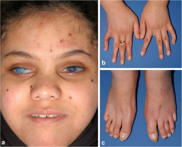
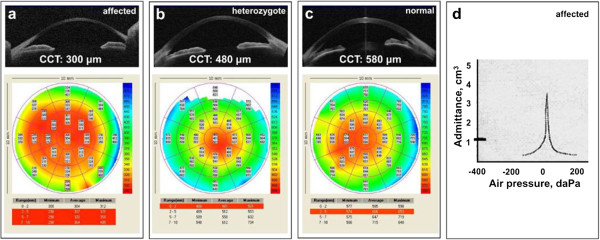
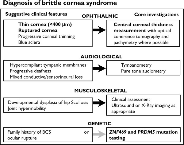
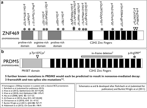
Brittle cornea syndrome (BCS) is an autosomal recessive disorder characterised by extreme corneal thinning and fragility. Corneal rupture can therefore occur either spontaneously or following minimal trauma in affected patients. Two genes, ZNF469 and PRDM5, have now been identified, in which causative pathogenic mutations collectively account for the condition in nearly all patients with BCS ascertained to date. Therefore, effective molecular diagnosis is now available for affected patients, and those at risk of being heterozygous carriers for BCS. We have previously identified mutations in ZNF469 in 14 families (in addition to 6 reported by others in the literature), and in PRDM5 in 8 families (with 1 further family now published by others). Clinical features include extreme corneal thinning with rupture, high myopia, blue sclerae, deafness of mixed aetiology with hypercompliant tympanic membranes, and variable skeletal manifestations. Corneal rupture may be the presenting feature of BCS, and it is possible that this may be incorrectly attributed to non-accidental injury. Mainstays of management include the prevention of ocular rupture by provision of protective polycarbonate spectacles, careful monitoring of visual and auditory function, and assessment for skeletal complications such as developmental dysplasia of the hip. Effective management depends upon appropriate identification of affected individuals, which may be challenging given the phenotypic overlap of BCS with other connective tissue disorders.


Figures

Figure 1
Figure 1
Clinical appearance of patient with BCS. This patient has a homozygous ZNF469 c.6444delG mutation (patient P3, Rohrbach et al. [6]) and is pictured at 18 years of age: a) face, b) hands and c) feet. Note extensive corneal scarring bilaterally and blue sclerae. Her facial appearance is reminiscent of several other patients with BCS, with a short nose, but otherwise unremarkable morphology. Several naevi are present over her cheeks and the lesions on her forehead are small scabs. In the hands, bilateral clinodactyly is seen, and in the feet, pes planus and scars of previous surgical management of hallux valgus.
Figure 2
Figure 2
Investigations in BCS. (a) Ocular optical coherence tomography (OCT) and pachymetry of a 32 year old patient with homozygous mutation in PRDM5 (deletion of exons 11–16; IV:4 of family BCS-001, Burkitt Wright et al. [3]). Note extreme thinning, particularly of the central cornea (300 μm) but even the maximum thickness at the periphery is only 330–380 μm. (b) Ocular OCT and pachymetry of a 31 year old patient with heterozygous mutation in PRDM5 (IV:8, BCS-001). Note only mild corneal thinning on OCT, with mean CCT measurement of 480 μm, increasing to 580–620 μm at the periphery. (c) Ocular OCT and pachymetry of a normal eye for comparison with a) and b), showing CCT of 580 μm and peripheral thickness of 650–750 μm. (d) Tympanogram of BCS patient (IV:4, BCS-001): a Type Ad curve is observed, demonstrating normal middle ear pressure but hypercompliance of the tympanic membrane. The volume by which the eardrum is displaced when a pulse of pressure is delivered to it is represented by the curve (~3.5 cm3), whilst the marker on the left demonstrates the degree of compliance observed (~1 cm3) for a normal tympanic membrane.
Figure 3
Figure 3
Diagnostic algorithm for patients with suspected brittle cornea syndrome.
Figure 4
Figure 4
Mutational spectrum in patients diagnosed with BCS. Note a greater proportion of mutations in (a) ZNF469 than (b) PRDM5, and a wide spectrum of mutations consistent with loss-of-function alleles. Articles in which the mutations are described are listed below [23,24].

Similar articles

Cited by

References

    1. Abu A, Frydman M, Marek D, Pras E, Nir U, Reznik-Wolf H, Pras E. Deleterious mutations in the Zinc-Finger 469 gene cause brittle cornea syndrome. Am J Hum Genet. 2008;82:1217–22. doi: 10.1016/j.ajhg.2008.04.001. - DOI - PMC - PubMed
    1. Christensen AE, Knappskog PM, Midtbø M, Gjesdal CG, Mengel-From J, Morling N, Rødahl E, Boman H. Brittle cornea syndrome associated with a missense mutation in the zinc-finger 469 gene. Invest Ophthalmol Vis Sci. 2010;51:47–52. doi: 10.1167/iovs.09-4251. - DOI - PubMed
    1. Burkitt Wright EM, Spencer HL, Daly SB, Manson FD, Zeef LA, Urquhart J, Zoppi N, Bonshek R, Tosounidis I, Mohan M, Madden C, Dodds A, Chandler KE, Banka S, Au L, Clayton-Smith J, Khan N, Biesecker LG, Wilson M, Rohrbach M, Colombi M, Giunta C, Black GC. Mutations in PRDM5 in brittle cornea syndrome identify a pathway regulating extracellular matrix development and maintenance. Am J Hum Genet. 2011;88:767–77. doi: 10.1016/j.ajhg.2011.05.007. - DOI - PMC - PubMed
    1. Aldahmesh MA, Mohamed JY, Alkuraya FS. A novel mutation in PRDM5 in brittle cornea syndrome. Clin Genet. 2012;81:198–9. doi: 10.1111/j.1399-0004.2011.01808.x. - DOI - PubMed
    1. Khan AO, Aldahmesh MA, Alkuraya FS. Brittle cornea without clinically-evident extraocular findings in an adult harboring a novel homozygous ZNF469 mutation. Ophthalmic Genet. 2012;33:257–9. doi: 10.3109/13816810.2012.670362. - DOI - PubMed

Publication types

Supplementary concepts