Review
. 2013 May;8(5):607-20.
doi: 10.2217/fmb.13.30.

Genetic diversity in the oral pathogen Porphyromonas gingivalis: molecular mechanisms and biological consequences

Affiliations
Review

Genetic diversity in the oral pathogen Porphyromonas gingivalis: molecular mechanisms and biological consequences

Gena D Tribble et al. Future Microbiol. 2013 May.

Abstract

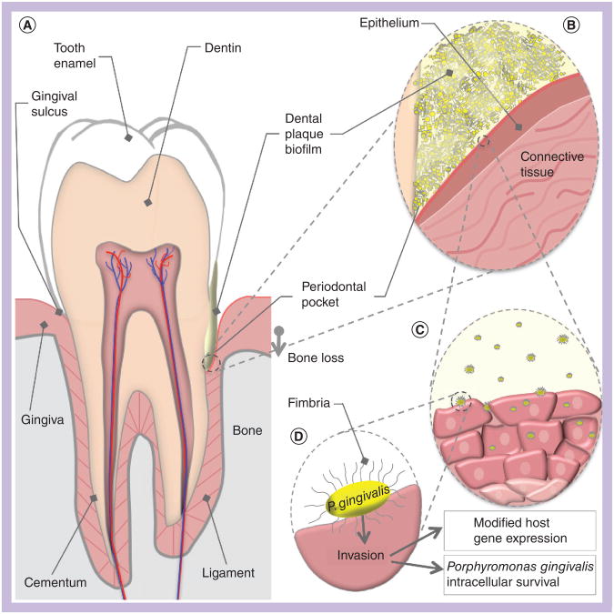
Porphyromonas gingivalis is a Gram-negative anaerobic bacterium that colonizes the human oral cavity. It is implicated in the development of periodontitis, a chronic periodontal disease affecting half of the adult population in the USA. To survive in the oral cavity, these bacteria must colonize dental plaque biofilms in competition with other bacterial species. Long-term survival requires P. gingivalis to evade host immune responses, while simultaneously adapting to the changing physiology of the host and to alterations in the plaque biofilm. In reflection of this highly variable niche, P. gingivalis is a genetically diverse species and in this review the authors summarize genetic diversity as it relates to pathogenicity in P. gingivalis. Recent studies revealing a variety of mechanisms by which adaptive changes in genetic content can occur are also reviewed. Understanding the genetic plasticity of P. gingivalis will provide a better framework for understanding the host-microbe interactions associated with periodontal disease.


Figures

Figure 1
Figure 1. Porphyromonas gingivalis host environment and interactions
(A) A cross-sectional schematic diagram of a human tooth. The periodontium is composed of the gingiva (pink) and the alveolar bone (grey). The left side represents periodontal health, while the right represents the changes that occur in periodontitis. Dental plaque is represented in yellow, in contact with the gingiva and tooth surfaces. Development of a chronic host inflammatory response ultimately results in a loss of connection between the gingiva and the root surface, creating a periodontal pocket. Bone is also resorbed as the bacteria advance down the surface of the tooth. (B) A magnified view of the periodontal pocket. The biofilm (yellow) is composed of many species of bacteria, of which Porphyromonas gingivalis is a small portion. P. gingivalis in the biofilm comes into direct contact with gingival epithelium. Secretion of P. gingivalis gingipains aids in the destruction of host antibodies and complement, and contributes to localized tissue destruction. (C) A number of P. gingivalis cells may leave the biofilm community and invade host cells. Internalized bacteria move from cell to cell into the deeper layers of the connective tissue. (D) Major fimbriae on the bacterial cell surface act as adhesins for host integrin receptors, and mediate internalization of the bacteria into the host cell cytoplasm. Postinvasion host cell gene expression is significantly modified; for example, reductions in cytokine production interferes with neutrophil recruitment to the periodontal pocket. The ability of P. gingivalis to modify local host responses to the biofilm flora results in the skewed immune response that drives periodontitis progression.
Figure 2
Figure 2. Transmission electron micrograph of Porphyromonas gingivalis strains 33277, 53977, 49417 and W83
Porphyromonas gingivalis cells from 2 days' growth on blood agar stained with 2% phosphotungstic acid, shown at 100,000× magnification. Membrane blebs are indicated with black arrows, major fimbriae with white arrows. (A) Strain 33277 (fimA type I). (B) Strain 53977 (fimA type II). (C) Strain 49417 (fimA type III). (D) Strain W83 (fimA type IV) does not display any fimbriae nor any obvious evidence of membrane blebbing. Scale bar: 100 nm. Sample preparation: Jennifer Kerr.
Figure 3
Figure 3. Examples of genomic differences between strains W83, 33277 and TDC60
The genome sequence of strain W83 was screened against strains 33277 and TDC60 to identify regions of conserved (dark blue) and divergent (grey) sequence. The upper alignment represents a region primarily encoding lipoproteins. The lower alignment demonstrates highly diverged regions flanked by conserved mobile elements. The genomic comparison was performed using CLC Genomics Workbench V4, using the internal BLAST function (Mask low-complexity; Expect 1; Match 1, Mismatch -2; Word size 30). The open reading frame annotations are from strain W83: yellow arrows represent coding sequence and light blue arrows represent mobile elements. ‘A’ denotes genes that are highly divergent between all three strains and are thus ‘strain-specific’. In the upper panel, ragAB encodes an outer membrane lipoprotein and PG0183 encodes a putative lipoprotein. ‘B’ denotes changes in DNA sequence in one of the three strains, TDC60, which result in limited changes in amino acid content. PG0181 and PG0182 encode putative lipoproteins; the genetic differences in strain TDC60 result in changes to 15 and 32 amino acids, respectively. ‘C’ denotes a mobile element that is found only in strain W83, ISPg4 to the left; and a mobile element found in all three strains, ISPg1 to the right. ‘D’ denotes genetic variation in the promoter region for PG0179. The promoter region is conserved between W83 and 33277, and divergent in TDC60. The two points of genetic variance begin 147 and 350 bp upstream of PG0179, and represent 28 and 25% genetic divergence over 127 and 385 bp, respectively. ‘E’ denotes conserved mobile elements flanking strain-specific genes. Two copies of the conserved ISPg1, left and middle, and one copy of ISPg2, right, flank regions containing W83-specifc genes encoding primarily hypothetical proteins. PG0861 is predicted to be a helicase and PG0862 a restriction endonuclease, suggesting that this region may encode functions associated with DNA transfer.
Figure 4
Figure 4. Mechanisms of DNA transfer in Porphyromonas gingivalis
Porphyromonas gingivalis resides in dental plaque biofilms in the presence of diverse bacterial species and multiple strains of P. gingivalis may coexist within the same biofilm community. (A)P. gingivalis biofilms have been shown to release eDNA (purple) into the biofilm matrix. This DNA can be taken up by other strains of P. gingivalis by natural transformation. This process requires the comF gene in the recipient strain; comF proteins provide the molecular motor to bring DNA across the bacterial membrane. DNA that is homologous to the host cell genome may be integrated into the chromosome by homologous recombination. (B) Some strains of P. gingivalis contain conjugative transposons that are normally integrated into the chromosome (black line), but may excise and form a circular intermediate prior to conjugative transfer to a new host cell (black circle). Conjugative transposon cTnPg1 has been shown to transfer between P. gingivalis strains, is able to transfer into other closely related species such as Prevotella (D), and can also mobilize plasmids and other genetic elements between species (dark blue circles). (C) Conjugation can also mediate the transfer of chromosomal DNA between strains of P. gingivalis. This process is dependent on conjugative transposons or other traAQ-containing elements present in the donor strain and homologous recombination in the recipient. Allelic replacement of recipient DNA (red) with donor DNA (blue line) is mediated by homologous recombination. Finally, insertion elements (light blue boxes) are present in P. gingivalis and are capable of transposing into genes or their promoter regions, thus influencing expression. They are also likely responsible for the large scale genomic rearrangements that can be detected by comparison of multiple genome sequences. (D) cTnPg1 can be transferred to the closely related genus Prevotella. eDNA: Extracellular DNA.

References

    1. Eke PI, Dye BA, Wei L, Thornton-Evans GO, Genco RJ. CDC periodontal disease surveillance workgroup Prevalence of periodontitis in adults in the United States: 2009 and 2010. J Dent Res. 2012;91(10):914–920. - PubMed
    1. Lamont RJ, Jenkinson HF. Subgingival colonization by Porphyromonas gingivalis. Oral Microbiol Immunol. 2000;15(6):341–349. - PubMed
    1. Kuboniwa M, Lamont RJ. Subgingival biofilm formation. Periodontology 2000. 2010;52(1):38–52. - PMC - PubMed
    1. Anerud A, Löe H, Boysen H, Smith M. The natural history of periodontal disease in man. Changes in gingival health and oral hygiene before 40 years of age. J Periodontol Res. 1979;14(6):526–540. - PubMed
    1. Otomo-Corgel J, Pucher JJ, Rethman MP, Reynolds MA. State of the science: chronic periodontitis and systemic health. J Evid Based Dent Pract. 2012;12(Suppl. 3):S20–S28. - PubMed

Website

    1. Porphyromonas gingivalis MLST database. http://pubmlst.org/pgingivalis.

Publication types