

Functional organization of neuronal and humoral signals regulating feeding behavior
- PMID: 23642202
- PMCID: PMC3991304
- DOI: 10.1146/annurev-nutr-071812-161125
Functional organization of neuronal and humoral signals regulating feeding behavior
Abstract
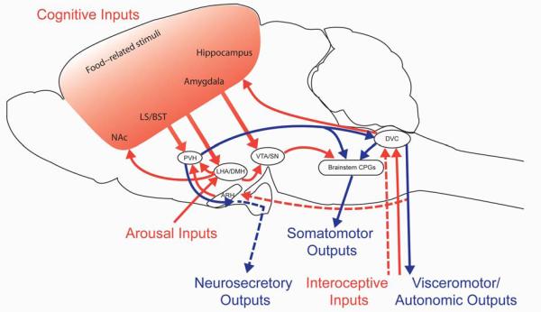
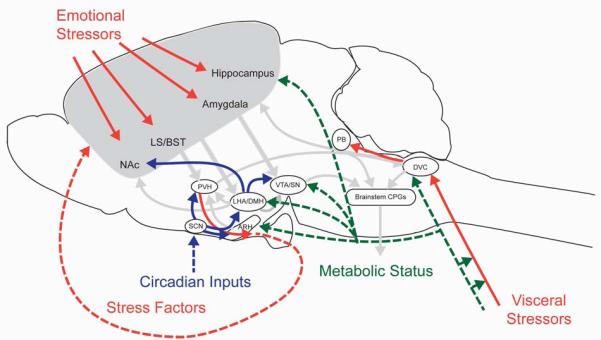
Energy homeostasis--ensuring that energy availability matches energy requirements--is essential for survival. One way that energy balance is achieved is through coordinated action of neural and neuroendocrine feeding circuits, which promote energy intake when energy supply is limited. Feeding behavior engages multiple somatic and visceral tissues distributed throughout the body--contraction of skeletal and smooth muscles in the head and along the upper digestive tract required to consume and digest food, as well as stimulation of endocrine and exocrine secretions from a wide range of organs. Accordingly, neurons that contribute to feeding behaviors are localized to central, peripheral, and enteric nervous systems. To promote energy balance, feeding circuits must be able to identify and respond to energy requirements, as well as the amount of energy available from internal and external sources, and then direct appropriate coordinated responses throughout the body.
Figures
Similar articles
-
Peripheral signals conveying metabolic information to the brain: short-term and long-term regulation of food intake and energy homeostasis.Exp Biol Med (Maywood). 2001 Dec;226(11):963-77. doi: 10.1177/153537020122601102. Exp Biol Med (Maywood). 2001. PMID: 11743131 Review.
-
Plasticity of central autonomic neural circuits in diabetes.Biochim Biophys Acta. 2009 May;1792(5):423-31. doi: 10.1016/j.bbadis.2008.12.001. Epub 2008 Dec 9. Biochim Biophys Acta. 2009. PMID: 19110053 Free PMC article. Review.
-
Synaptic plasticity in neuronal circuits regulating energy balance.Nat Neurosci. 2012 Oct;15(10):1336-42. doi: 10.1038/nn.3219. Epub 2012 Sep 25. Nat Neurosci. 2012. PMID: 23007188 Review.
-
Feeding behavior in mammals including humans.Ann N Y Acad Sci. 2009 Apr;1163:221-32. doi: 10.1111/j.1749-6632.2008.03627.x. Ann N Y Acad Sci. 2009. PMID: 19456343 Review.
-
Role of sensory input in the control of food intake.J Auton Nerv Syst. 1984 May-Jun;10(3-4):347-58. doi: 10.1016/0165-1838(84)90032-8. J Auton Nerv Syst. 1984. PMID: 6481094
Cited by
-
NEUROSCIENCE. A satiating signal.Science. 2016 Mar 18;351(6279):1268-9. doi: 10.1126/science.aaf5216. Epub 2016 Mar 17. Science. 2016. PMID: 26989239 Free PMC article. No abstract available.
-
Sensing of triacylglycerol in the gut: different mechanisms for fatty acids and 2-monoacylglycerol.J Physiol. 2015 Apr 15;593(8):2097-109. doi: 10.1113/jphysiol.2014.285635. Epub 2015 Feb 9. J Physiol. 2015. PMID: 25639597 Free PMC article.
-
Central insulin signaling modulates hypothalamus-pituitary-adrenal axis responsiveness.Mol Metab. 2014 Dec 10;4(2):83-92. doi: 10.1016/j.molmet.2014.12.001. eCollection 2015 Feb. Mol Metab. 2014. PMID: 25685696 Free PMC article.
-
Metabolic vs. hedonic obesity: a conceptual distinction and its clinical implications.Obes Rev. 2015 Mar;16(3):234-47. doi: 10.1111/obr.12246. Epub 2015 Jan 14. Obes Rev. 2015. PMID: 25588316 Free PMC article. Review.
-
Incretins and amylin: neuroendocrine communication between the gut, pancreas, and brain in control of food intake and blood glucose.Annu Rev Nutr. 2014;34:237-60. doi: 10.1146/annurev-nutr-071812-161201. Epub 2014 Apr 10. Annu Rev Nutr. 2014. PMID: 24819325 Free PMC article.
References
-
- Banks WA, Kastin AJ, Huang W, Jaspan JB, Maness LM. Leptin enters the brain by a saturable system independent of insulin. Peptides. 1996;17:305–11. - PubMed
-
- Banks WA, Tschop M, Robinson SM, Heiman ML. Extent and direction of ghrelin transport across the blood-brain barrier is determined by its unique primary structure. J Pharmacol Exp Ther. 2002;302:822–7. - PubMed
Publication types
MeSH terms
Grants and funding
LinkOut - more resources
Full Text Sources
Other Literature Sources
