Review
. 2013:82:81-118.
doi: 10.1146/annurev-biochem-072711-165700.

Readout of epigenetic modifications

Affiliations
Review

Readout of epigenetic modifications

Dinshaw J Patel et al. Annu Rev Biochem. 2013.

Abstract

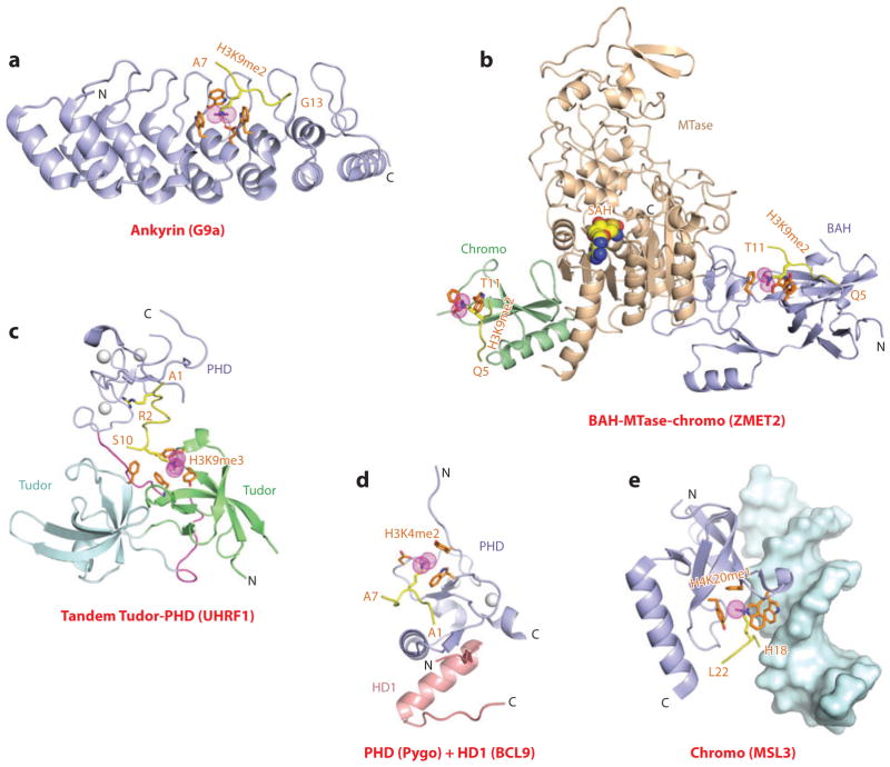
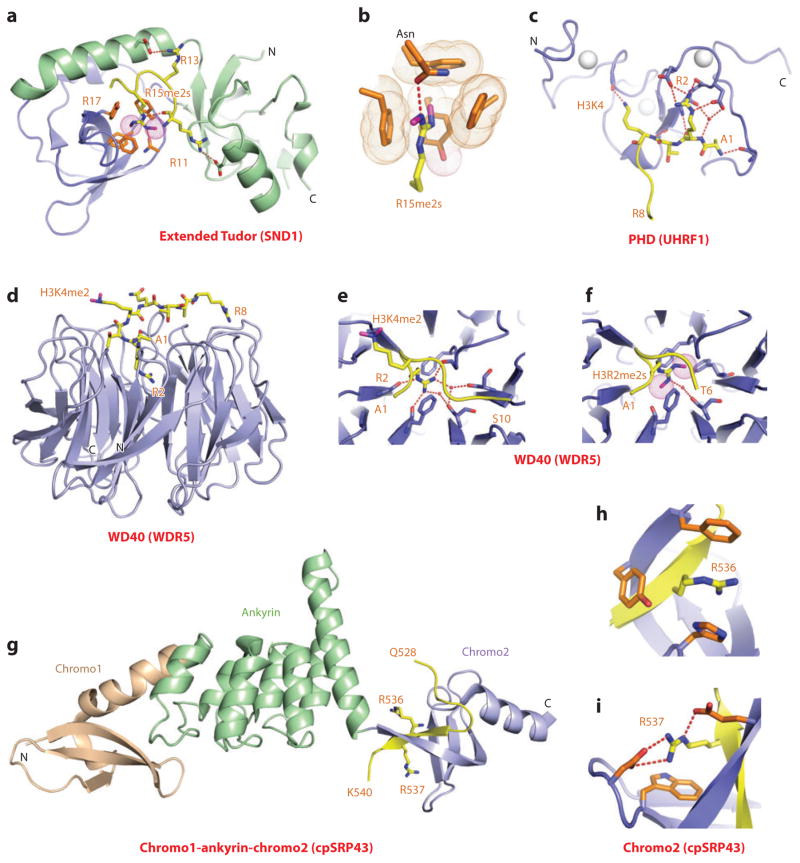
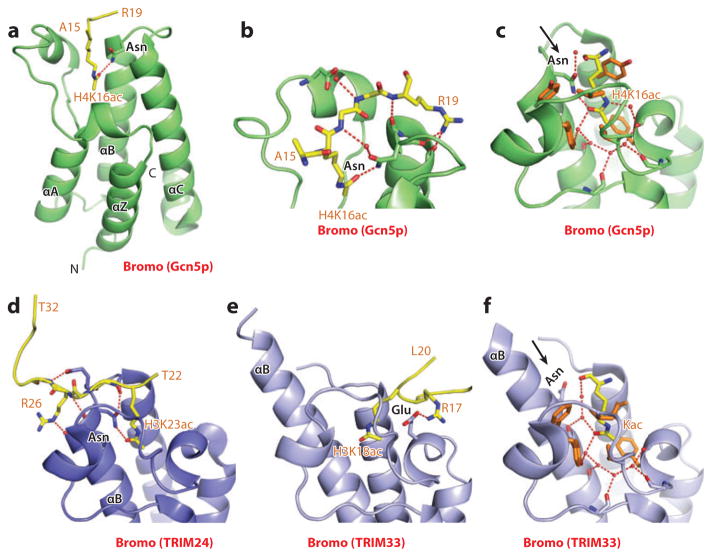
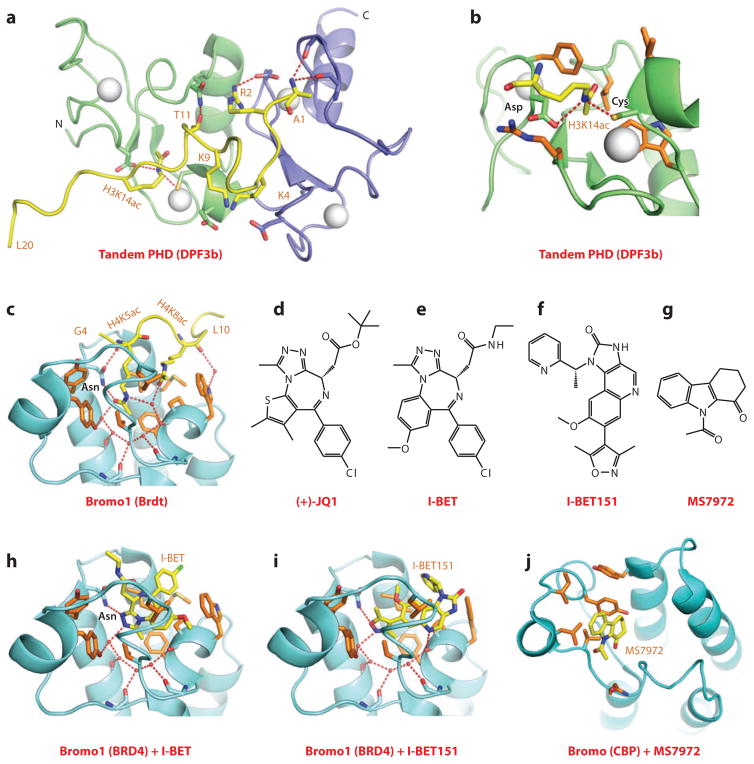
This review focuses on a structure-based analysis of histone posttranslational modification (PTM) readout, where the PTMs serve as docking sites for reader modules as part of larger complexes displaying chromatin modifier and remodeling activities, with the capacity to alter chromatin architecture and templated processes. Individual topics addressed include the diversity of reader-binding pocket architectures and common principles underlying readout of methyl-lysine and methyl-arginine marks, their unmodified counterparts, as well as acetyl-lysine and phosphoserine marks. The review also discusses the impact of multivalent readout of combinations of PTMs localized at specific genomic sites by linked binding modules on processes ranging from gene transcription to repair. Additional topics include cross talk between histone PTMs, histone mimics, epigenetic-based diseases, and drug-based therapeutic intervention. The review ends by highlighting new initiatives and advances, as well as future challenges, toward the promise of enhancing our structural and mechanistic understanding of the readout of histone PTMs at the nucleosomal level.


Figures

Figure 1
Figure 1
Structures of single “Royal family” modules bound to methyl-lysine-containing histone peptides. (a) The 2.4-Å crystal structure of the complex of the HP1 chromodomain bound to the H3(1–15)K9me3 peptide, Protein Data Bank (PDB) reference 1KNE. The bound K9me3-containing H3 peptide can be traced from Gln5 to Ser10 (Q5 to S10). (b) Details of the antiparallel alignment of the β-strand of the bound H3K9me3-containing peptide sandwiched between the β-strands of the HP1 chromodomain, resulting in generation of a three-stranded antiparallel β-pleated sheet on formation of the complex. (c) The 1.85-Å crystal structure of the complex of the PHF1 Tudor domain bound to the H3(31–40)K36me3 peptide complex (PDB: 4HCZ). The bound K36me3-containing H3 peptide can be traced from Ser31 to Arg40 (S31 to R40). (d) The 1.5-Å crystal structure of the complex of the Brf1 PWWP domain bound to the H3(22–42)K36me3 peptide complex (PDB: 2X4W). The bound K36me3-containing H3 peptide can be traced from Ser28 to Arg40 (S28 to R40). Abbreviations: C, C terminus; N, N terminus.
Figure 2
Figure 2
Structures of the PHD finger (BPTF) and BAH domain (mammalian ORC1) bound to methyl-lysine-containing histone peptides. (a) The 2.0-Å crystal structure of the complex of the BPTF PHD finger bound to the H3(1–15)K4me3 peptide, Protein Data Bank (PDB) reference 2F6J. The PHD finger (as part of a PHD-bromo cassette) in a ribbon representation is shown (blue), with two stabilizing bound Zn ions (silver balls). The bound peptide from Ala1 to Thr6 (A1 to T6) is in yellow with the trimethyl group of Kme3 shown as dotted magenta balls. The residues forming the aromatic-lined cage pocket are orange. (b) Positioning of Arg2 (R2) and Lys4me3 (K4me3) side chains in the adjacent open surface pockets (surface groove mode), separated by the indole ring of an invariant Trp in the complex. The PHD finger and peptide in the complex are shown in surface and space-filling representations, respectively. (c) Details showing the antiparallel alignment of the β-strands of the bound H3K4me3-containing peptide and PHD finger, resulting in generation of an antiparallel β-pleated sheet on formation of the complex. Note that the positively charged N terminus is anchored in its own pocket. (d) Positioning of the K4me3 group within the aromatic-lined cage in the complex. (e) Positioning of the K4me2 group into an engineered pocket containing a Glu residue that replaced the Tyr residue in panel d (PDB: 2RIJ). (f) The 1.43-Å crystal structure of the complex of the PHD finger of BHC80 bound to the H3(1–10) peptide (PDB: 2PUY). The bound H3 peptide can be traced from Ala1 to Ser10 (A1 to S10). (g) The 1.95-Å crystal structure of the complex of the mouse ORC1 BAH domain bound to the H4(14–25)K20me2 peptide (PDB: 4DOW). The bound K20me2-containing H4 peptide can be traced from Gly14 to Arg23 (G14 to R23). (h) Details of the alignment of the K20me2-containing H4 peptide from G14 to R23 positioned on the mouse ORC1 BAH domain in the complex. The dimethylammonium group of H4K20 inserts into an aromatic-lined pocket in the BAH domain. Abbreviations: C, C terminus; N, N terminus.
Figure 3
Figure 3
Structures of malignant brain tumor (MBT, i.e., L3MBTL1) and ADD (ATRX) domains bound to methyl-lysine-containing histone peptides. (a) The 1.66-Å crystal structure of the complex of L3MBTL1 bound to the H1(22–26)K26me2 peptide, Protein Data Bank (PDB) reference 2RHI. A C-terminal peptide from an adjacent L3MBTL1 inserts proline 523 (P523) into the aromatic-lined pocket of the MBT domain 1 (pink). The dimethylammonium group of bound K26me2 inserts into the aromatic-lined pocket of the MBT domain 2 (blue), with the K26me2-containing H1 peptide traced from Thr24 to Lys26me2 (T24 to K26me2). A polyethylene glycol (PEG) molecule inserts into the aromatic-lined pocket of the MBT domain 3 (green). (b) Details of insertion (cavity insertion mode) of the dimethylammonium group of bound K26me2 into the aromatic-lined pocket of the MBT domain 2. (c) Details of insertion of the P523 from an adjacent L3MBTL1 into the aromatic-lined pocket of the MBT domain 1. This pocket is shallower than the one shown in panel b. (d) Chemical formula of UNC669, a pyrrolidine-containing small molecule. (e) Details of insertion of UNC669 into the aromatic-lined pocket of MBT domain 2 based on the 2.55-Å crystal structure of L3MBTL1 bound to UNC669 (PDB: 3P8H). (f) The 1.6-Å crystal structure of the complex of the ADD domain of ATRX bound to the H3(1–15)K9me3 peptide (PDB: 3QLA). The GATA-like and PHD fingers are green and blue, respectively. Bound Zn ions are shown (silver balls). The K9me3-containing H3 peptide is traced from A1 to S10. (g) Details of the intermolecular contacts involving the bound K9me3-containing H3 peptide in the complex, with the bound peptide traced from Ala1 to Ser10 (A1 to S10). (h) A surface and space-filling representation of the surface complementarity between K9me3 and the walls of the composite pocket lined by the GATA-like and PHD finger domains in the complex. (i) Ribbon and stick representation of K9me3 positioned to interact with the GATA-like and PHD finger domains in the complex. Abbreviations: C, C terminus; N, N terminus.
Figure 4
Figure 4
Structures of tandem Royal family modules bound to methyl-lysine-containing histone peptides. (a) The 2.4-Å crystal structure of the complex of double chromo domains of human CHD1 proteins bound to the H3(1–19)K4me3 peptide, Protein Data Bank (PDB) reference 2B2W. Tudor domains 1 and 2 are shown (blue and green, respectively) with the connecting helix-turn-helix linker (pink). The bound K4me3-containing H3 peptide can be traced from Ala1 to Gln5 (A1 to Q5). (b) The 1.7-Å crystal structure of the complex of tandem Tudor domains of 53BP1 bound to the H4(15–24)K20me2 peptide (PDB: 2IG0). Tudor domains 1 and 2 are blue and green. The bound K20me2-containing H4 peptide can be traced for the Arg19-Lys20me2 (R19-K20me2) step. (c) The 2.1-Å crystal structure of the complex of tandem Tudor domains of JMJD2A bound to the H3(1–10)K4me3 peptide (PDB: 2GFA). Individual Tudor domains are blue and green. The bound K4me3-containing peptide can be traced from Ala1 to Ala7 (A1 to A7). (d) The 1.26-Å crystal structure of the complex of tandem Tudor domains of Sgf29 bound to the H3(1–11)K4me3 peptide (PDB: 3MEA). Tudor domains 1 and 2 are blue and green. The bound K4me3-containing peptide can be traced from Ala1 to Lys4me3 (A1 to K4me3). (e) The 2.7-Å crystal structure of the complex of tandem Tudor domains of the Arabidopsis thaliana SHH1 protein bound to the H3(1–15)K9me2 peptide (PDB: 4IUT). A bound zinc ion is shown (silver ball). Tudor domains 1 and 2 are blue and green. The bound K9me2-containing H3 peptide can be traced from Thr3 to Ser10 (T3 to S10). Abbreviations: C, C terminus; N, N terminus.
Figure 5
Figure 5
Structures of linked binding and accessory modules involved in multivalent readout of methyl-lysine-containing histone peptides. (a) The 2.99-Å crystal structure of the complex of the ankyrin repeats of G9a bound to the H3(1–15)K9me2 peptide, Protein Data Bank (PDB) reference 3B95. The aromatic-lined binding pocket bound by K9me2 is positioned between the fourth and fifth ankyrin repeats of G9a. The bound K9me2-containing H3 peptide can be traced from Ala7 to Gly13 (A7 to G13). (b) A superposed view of the crystal structures of the complex of the BAH domain–methyltransferase (MTase) domain–chromodomain of maize ZMET2 bound to the H3(1–32)K9me2 peptide (2.7 Å, bound to BAH domain) (PDB: 4FT4) and bound to the H3(1–15)K9me2 peptide (3.2 Å, bound to chromodomain) (PDB: 4FT2). The chromodomain, BAH, and methyltransferase domains are green, blue, and beige, respectively. The bound K9me2-containing H3 peptides can be traced from Gln5 to Thr11 (Q5 to T11) when bound to both the BAH and chromodomains. (c) The 2.9-Å crystal structure of the complex of the tandem Tudor-PHD finger cassette of UHRF1 bound to the H3(1–13)K9me3 peptide (PDB: 3ASK). The tandem Tudor domains are shown in the lower segment (cyan and green), and the PHD finger is shown in the upper segment (blue). The bound H3(1–13)K9me3-containing peptide can be traced from Ala1 to Ser10 (A1 to S10). (d) The 1.7-Å crystal structure of the ternary complex of the Pygopus PHD finger (blue) bound to the H3(1–7)K4me2 peptide in the presence of the homology domain 1 (HD1) domain of BCL9 (pink) (PDB: 2VPE). The bound K4me2-containing H3 peptide can be traced from Ala1 to Ala7 (A1 to A7). (e) The 2.35-Å crystal structure of the complex of MSL3 chromodomain bound to the H4(9–31)K20me1 peptide in the presence of duplex DNA (surface representation) (PDB: 3OA6). The bound K20me1-containing H4 peptide can be traced from His18 to Leu22 (H18 to L22). Abbreviations: C, C terminus; N, N terminus.
Figure 6
Figure 6
Structures of expanded and paired modules bound to methyl-arginine and unmodified arginine-containing peptides. (a) The 2.8-Å crystal structure of the complex of the extended Tudor module of SND1 bound to R15me2s-containing N-terminal PIWI peptide, Protein Data Bank (PDB) reference 3NTI. The core fold of the Tudor domain is shown in blue, and the extensions are shown in green. The R15me2s-containing N-terminal PIWI peptide can be traced from Arg11 to Arg17 (R11 to R17). (b) Positioning of R15me2s in the aromatic-lined cage pocket of the Tudor domain in the SND1 complex. (c) The 1.8-Å crystal structure of the complex of the PHD finger of UHRF1 bound to the H3(1–9) peptide (PDB: 3SOU). The bound H3 peptide can be traced from Ala1 to Arg8 (A1 to R8). Note the network of hydrogen bonds involving Arg2 (R2) and residues on the PHD finger. (d) The 1.5-Å crystal structure of the complex of the WD40 motif of WDR5 bound to the H3(1–9)K4me2 peptide. The bound K4me2-containing peptide can be traced from Ala1 to Arg8 (A1 to R8) (PDB: 2H6N). (e) Intermolecular hydrogen-bonding interactions stabilizing insertion of Arg2 (R2) into the central channel of the WD40 motif in the H3(1–9)K4me2-WDR5 complex. (f) Insertion of symmetrical Arg2me2 (R2me2s) into the central channel of the WD40 motif in the H3(1–15)R2me2s-WDR5 complex solved at 1.9 Å (PDB: 4A7J). (g) The 3.18-Å crystal structure of the complex of the chromodomains and ankyrin repeats of Arabidopsis thaliana cpSRP43 bound to an Arg-Arg-Lys-Arg (RRKR)-containing peptide (PDB: 3UI2). The side chains of Arg536 (R536) and Arg537 (R537) of the bound RRKR-containing peptide from Gln528 to Lys540 (Q528-K540) are positioned in adjacent pockets at the interface between the fourth ankyrin repeat and the second chromodomain in the complex. (h) Position of Arg536 (R536) of the RRKR-containing peptide within an aromatic-lined cage pocket in the complex. (i) Positioning of Arg537 (R537) of the RRKR-containing peptide in a pocket lined by a Trp and two acidic side chains in the complex. Abbreviation: C, C terminus.
Figure 7
Figure 7
Structures of bromodomains bound to acetyl-lysine-containing histone peptides. (a) The 1.87-Å structure of the bromodomain of Gcn5p bound to the H4(15–29)K16ac peptide, Protein Data Bank (PDB) reference 1E6I. The peptide can be traced from Ala15 to Arg19 (A15 to R19). (b) Details of intermolecular recognition between the peptide backbone and loops that project from the bromodomain scaffold. (c) Details of intermolecular contacts in the binding pocket of the Gcn5p bromodomain-H4(15–29)K16ac complex. The acetyl-lysine side chain inserts into a hydrophobic pocket and is anchored through hydrogen bonding with an asparagine side chain. Also note the extensive use of water-mediated intermolecular recognition in the pocket. (d) The 1.9-Å structure of the bromodomain of TRIM24 bound to the H3(13–32)K23ac peptide (PDB: 3O34). The chain can be traced from Thr22 to Thr32 (T22 to T32), with the side chain of K23ac involved in canonical recognition within the binding pocket. Interactions involving the side chain of Arg26 (R26) of the bound peptide are also show in this panel. (e) The 2.7-Å structure of the bromodomain of TRIM33 bound to the H3(1–22)K9me3K14acK18ac peptide (PDB: 3U5O). The chain can be traced from Ala1 to Leu20 (A1 to L20), with the segment from Arg17 to Leu20 (R17 to L20) shown in this panel. The side chain of K18ac is involved in atypical recognition, in that it is not hydrogen bonded to an asparagine. Interactions involving the side chain of Arg17 (R17) of the bound peptide are also shown in this panel. (f) Details of intermolecular contacts (using the structure at 1.95-Å resolution) in the binding pocket of the TRIM33 bromodomain-H3(1–20)K9me3K14ac complex (PDB: 3U5N). The acetyl-lysine side chain inserts into a hydrophobic pocket and is anchored solely through water-mediated interactions. The extension of the α-helix B (αB) in TRIM33 results in rotation of the asparagine residue away from the pocket such that it is no longer involved in recognition of the Kac side chain. Abbreviations: αA, -C, α-helix A and C; C, C terminus; Kac, acetyl-lysine.
Figure 8
Figure 8
Structures of tandem PHD fingers (DPF3b) and bromodomains (BET family) bound to acetyl-lysine-containing histone peptides. (a) The NMR-based solution structure of the tandem PHD fingers of DPF3b bound to the H3(1–20)K14ac peptide, Protein Data Bank (PDB) reference 2KWJ. The first and second PHD fingers are shown as green and blue, respectively. The H3 peptide was traced from Ala1 to Leu20 (A1 to L20). Interactions involving the side chains of Arg2 (R2), Lys9 (K9), and Thr11 (T11) of the bound peptide are show in this panel. (b) Details of the alignment and interactions involving the side chain of K14ac with the first PHD finger in the DPF3b tandem PHD finger-H3(1–20)K14ac peptide complex. (c) The 2.37-Å structure of the first bromodomain of Brdt bound to the H4(1–20)K5acK8ac peptide (PDB: 2WP2). The bound peptide can be traced from Gly4 to Leu10 (G4 to L10). The side chain of K5ac inserts into the Kac-binding pocket, whereas the side chain of K8ac is on the outer rim of the pocket and buttresses the insertion of K5ac into the pocket. (d) Chemical formula of (+)-JQ1. (e) Chemical formula of I-BET. (f) Chemical formula of I-BET151. (g) Chemical formula of MS7972. (h) The 1.6-Å structure of the first bromodomain of BRD4 bound to the small-molecule inhibitor I-BET (PDB: 3P5O). I-BET inserts into the Kac-binding pocket and is anchored in place by shape complementarity and direct and water-mediated intermolecular contacts. (i) The 1.5-Å structure of the first bromodomain of BRD4 bound to the small-molecule inhibitor I-BET151 (PDB: 3ZYU). I-BET151 inserts into the Kac-binding pocket and is anchored in place by shape complementarity and direct and water-mediated intermolecular contacts. (j) The NMR-based solution structure of insertion of the small-molecule inhibitor MS7972 into the acetyl-lysine-binding pocket of the bromodomain of CBP. Abbreviation: N, N terminus.
Figure 9
Figure 9
Structures of 14-3-3 proteins bound to phosphoserine marks and inhibitors. (a) The 2.0-Å crystal structure of 14-3-3ζ bound to the H3(7–14)S10ph peptide, Protein Data Bank (PDB) reference 2C1N. The 14-3-3ζ protein in the complex adopts a symmetrical dimeric alignment, with individual monomers shown in blue and green. The bound peptide can be traced from Ala7 to Lys14 (A7 to K14). (b) Details of the alignment and hydrogen-bonding interactions involving the side chain of S10ph of the peptide in the H3(7–14)S10ph-14-3-3ζ complex. Also, note the pair of hydrogen bonds between the guanidinium group of Arg8 (R8) of the peptide and the carboxyl group of a Glu residue of the protein. (c) The 2.4-Å crystal structure of the ternary complex between Hd3a (pink), GF14c (blue), and the bound OsFD1(187–195)S192ph peptide (yellow) (PDB: 3AXY). The side chain can be traced from Arg189 to Phe195 (R189 to F195). (d) Details of the alignment and hydrogen-bonding interactions involving the side chain of phosphorylated Ser192 (S192ph) and the peptide in the ternary complex. (e) The chemical formula of FOBISIN101. (f) Details of interactions of the phosphate group of the cleaved pyridoxal-phosphate moiety of FOBISIN101 with 14-3-3ζ in the FOBISIN101-14-3-3ζ complex. Abbreviations: C, C terminus; N, N terminus.
Figure 10
Figure 10
Structures of BRCT (MCPH1) and BIR (Survivin) domains bound to phosphoserine/threonine/tyrosine-containing peptides. (a) The 1.5-Å crystal structure of the complex of the tandem BRCT domains of MCPH1 bound to di-γH2A.X(139–142)S139phY142ph peptide, Protein Data Bank (PDB) reference 3U3Z. (b) Details of the alignment and interactions involving the side chain of phosphorylated Ser139 (S139ph) and phosphorylated Tyr142 (Y142ph) (arrows) of the di-γH2A.X(139–142)S139phY142ph peptide with the first (blue) and second (green) BRCT domains of MCPH1. Note that Y142ph adopts two conformations in the structure of the complex. (c) The 2.4-Å crystal structure of the complex of the BIR domain of Survivin bound to the H3(1–15)T3ph peptide (PDB: 3UIG). The peptide chain can be traced from Ala1 to Gln5 (A1 to Q5). (d) Details of the alignment and interactions between the bound H3(1–15)T3ph peptide and the BIR domain of Survivin. Interactions involving the side chains of Arg2 (R2) and Lys4 (K4) are also shown in the panel. (e) Details of the alignment and interactions between the bound Smac/DIABLO(1–15) peptide and the BIR domain of Survivin (PDB: 3UIH). The peptide chain can be traced from Ala1 to Ile4 (A1 to I4). Abbreviations: C, C terminus; MCPH1, microcephalin 1; N, N terminus.
Figure 11
Figure 11
Structures and in vivo functional studies of PHD-bromo cassettes bound to methyl-lysine- and acetyl-lysine-containing histone peptides. (a) The 2.0-Å crystal structure of the complex of the PHD-bromo cassette of BPTF bound to the H3(1–15)K4me3 peptide, Protein Data Bank (PDB) reference 2F6Z. A separate 1.8-Å crystal structure of the bromodomain of BPTF bound to the H4(12–21)K16ac peptide was also solved (PDB: 3QZS), and that information was superimposed on the structure shown in this panel. The bound H3(1–15)K4me3-containing peptide can be traced from Ala1 to Thr6 (A1 to T6), and the bound H4(12–21)K16ac-containing peptide can be traced from Lys14 to Val21 (K14 to V21) in the complexes. (b) A glutathione S-transferase (GST) pull-down of modified nucleosomes containing semisynthetic histones produced by expressed protein ligation. Nucleosomes containing dual marks involving H4K12ac, H4K16ac, or H4K20ac in combination with H3K4me3 are pulled down with a resin-bound GST-BPTF PHD-bromo cassette and detected by autoradiography after native gel electrophoresis. (c) The 1.72-Å crystal structure of the MLL1 PHD-bromo cassette exhibits a cis linker proline in the free state (PDB: 3LQH). The PHD finger and bromodomains are blue and green, respectively. (d) Model of the MLL1 PHD-bromo cassette with a trans linker proline, with this alignment stabilized by the bound RRM domain of CyP33. (e) The 2.7-Å crystal structure of the complex of the PHD-bromo cassette of TRIM33 bound to the H3(1–22)K9me3K18ac peptide (PDB: 3U5O). The bound H3(1–22)K9me3K18ac peptide can be traced from Ala1 to Leu20 (A1 to L20) in the complex. Abbreviations: C, C terminal; CyP33, cyclophilin 33; MLL1, mixed-lineage leukemia 1 protein; N, N terminal; RRM, RNA recognition motif; Rel CPM, relative counts per minute.

References

    1. Luger K, Dechassa ML, Tremethick DJ. New insights into nucleosome and chromatin structure: an ordered state or a disordered affair? Nat Rev Mol Cell Biol. 2012;13:436–47. - PMC - PubMed
    1. Kornberg RD, Lorch Y. Twenty-five years of the nucleosome, fundamental particle of the eukaryote chromosome. Cell. 1999;98:285–94. - PubMed
    1. Kouzarides T. Chromatin modifications and their function. Cell. 2007;128:693–705. - PubMed
    1. Musselman CA, Lalonde ME, Cote J, Kutateladze TG. Perceiving the epigenetic landscape through histone readers. Nat Struct Mol Biol. 2012;19:1218–27. - PMC - PubMed
    1. Ruthenburg AJ, Li H, Patel DJ, Allis CD. Multivalent engagement of chromatin modifications by linked binding modules. Nat Rev Mol Cell Biol. 2007;8:983–94. - PMC - PubMed

Publication types