

Expression of matrix metalloproteinase-12 in aortic dissection
- PMID: 23642232
- PMCID: PMC3660235
- DOI: 10.1186/1471-2261-13-34
Expression of matrix metalloproteinase-12 in aortic dissection
Abstract
Background: Aortic dissection(AD) is an acute process of large blood vessels characterized by dangerous pathogenic conditions and high disability and high mortality. The pathogenesis of AD remains debated. Matrix metalloproteinase-12 (MMP-12) participates in many pathological processes such as abdominal aortic aneurysm, atherosclerosis, emphysema and cancer. However, this elastase has rarely been assessed in the presence of AD. The aim of the present study was to investigate the expression of MMP-12 in aortic tissue so as to offer a better understanding of the possible mechanisms of AD.
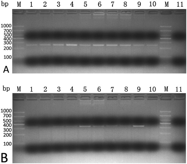
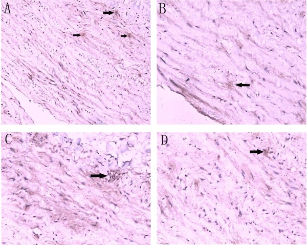
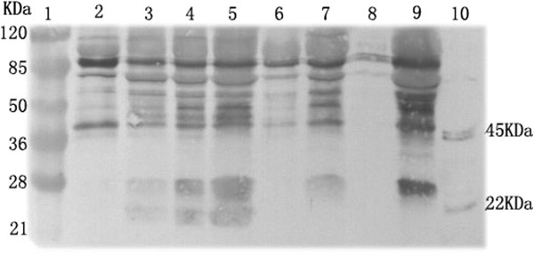
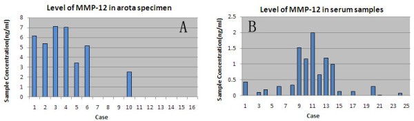
Methods: The protein expression levels of MMP-12 were analyzed and compared in aorta tissue and the blood serum samples by reverse transcription polymerase chain reaction(RT-PCR), Western blotting, immuno-histochemistry, fluorescence resonance energy transfer ( FRET ) activity assay and enzyme-linked immuno sorbent assay ( ELISA ), respectively. Ascending aorta tissue specimens were obtained from 12 patients with an acute Stanford A-dissection at the time of aortic replacement, and from 4 patients with coronary artery disease (CAD) undergoing coronary artery bypass surgery. Meanwhile, serum samples were harvested from 15 patients with an acute Stanford A-dissection and 10 healthy individuals who served as the control group.
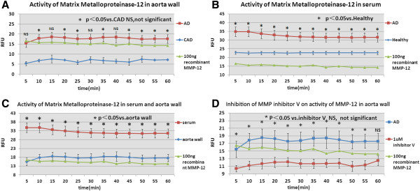
Results: MMP-12 activity could be detected in both AD and CAD groups, but the level in the AD group was higher than those in the CAD group (P < 0.05). MMP-12 proteolysis existed in both serum samples of the AD and healthy groups, and the activity level in the AD group was clearly higher than in the healthy group (P < 0.05). For AD patients, MMP-12 activity in serum was higher than in the aorta wall (P < 0.05). MMP-12 activity in the aortic wall tissue can be inhibited by MMP inhibitor v (P < 0.05).
Conclusion: The present study directly demonstrates that MMP-12 proteolytic activity exists within the aorta specimens and blood samples from aortic dissection patients. MMP-12 might be of potential relevance as a clinically diagnostic tool and therapeutic target in vascular injury and repair.
Figures
Similar articles
-
Differential expression of elastin assembly genes in patients with Stanford Type A aortic dissection using microarray analysis.J Vasc Surg. 2011 Apr;53(4):1071-1078.e2. doi: 10.1016/j.jvs.2010.11.035. Epub 2011 Jan 26. J Vasc Surg. 2011. PMID: 21276682
-
Plasma levels of metalloproteinases-9 and -2 in the acute and subacute phases of type A and type B aortic dissection.J Cardiovasc Med (Hagerstown). 2006 May;7(5):307-15. doi: 10.2459/01.JCM.0000223251.26988.c5. J Cardiovasc Med (Hagerstown). 2006. PMID: 16645407
-
Osteopontin expression and its possible functions in the aortic disorders and coronary artery disease.Rev Bras Cir Cardiovasc. 2011 Apr-Jun;26(2):173-82. doi: 10.1590/s0102-76382011000200006. Rev Bras Cir Cardiovasc. 2011. PMID: 21894406
-
Matrix metalloproteinases in aortic dissection.Vascul Pharmacol. 2024 Sep;156:107420. doi: 10.1016/j.vph.2024.107420. Epub 2024 Aug 23. Vascul Pharmacol. 2024. PMID: 39182633 Review.
-
Stanford-A acute aortic dissection, inflammation, and metalloproteinases: a review.Ann Med. 2015;47(6):441-6. doi: 10.3109/07853890.2015.1073346. Epub 2015 Sep 4. Ann Med. 2015. PMID: 26339779 Review.
Cited by
-
Expression of matrix metalloproteinases-12 in ST-segment elevation myocardial infarction: A case-control study.Medicine (Baltimore). 2017 Oct;96(40):e8035. doi: 10.1097/MD.0000000000008035. Medicine (Baltimore). 2017. PMID: 28984758 Free PMC article.
-
Differentially expressed genes and canonical pathways in the ascending thoracic aortic aneurysm - The Tampere Vascular Study.Sci Rep. 2017 Sep 21;7(1):12127. doi: 10.1038/s41598-017-12421-4. Sci Rep. 2017. PMID: 28935963 Free PMC article.
-
Tracking an Elusive Killer: State of the Art of Molecular-Genetic Knowledge and Laboratory Role in Diagnosis and Risk Stratification of Thoracic Aortic Aneurysm and Dissection.Diagnostics (Basel). 2022 Jul 22;12(8):1785. doi: 10.3390/diagnostics12081785. Diagnostics (Basel). 2022. PMID: 35892496 Free PMC article. Review.
-
Platelets at the Vessel Wall in Non-Thrombotic Disease.Circ Res. 2023 Mar 17;132(6):775-790. doi: 10.1161/CIRCRESAHA.122.321566. Epub 2023 Mar 16. Circ Res. 2023. PMID: 36927182 Free PMC article. Review.
-
CXCR6 Mediates Pressure Overload-Induced Aortic Stiffness by Increasing Macrophage Recruitment and Reducing Exosome-miRNA29b.J Cardiovasc Transl Res. 2023 Apr;16(2):271-286. doi: 10.1007/s12265-022-10304-2. Epub 2022 Aug 26. J Cardiovasc Transl Res. 2023. PMID: 36018423
References
-
- National Center for Health Statistics. Centers for Disease Control and Prevention. United States Department of Health and Human Services. United States: Deaths, percent of total deaths and death rates for the 15 leading causes of death in 10-year age groups by race and sex. 2005. Available at: http://www.cdc.gov/nchs/data/dvs/LCWK2_2001.pdf. Accessed May 3.
-
- Kung HC, Hoyert DL, Xu JQ, Murphy SL. Deaths: final data for 2005. Natl Vital Stat Rep. 2008;56:33. - PubMed
-
- Hiratzka LF, Bakris GL, Beckman JA. et al.American College of Cardiology Foundation/American Heart Association Task Force on Practice Guidelines; American Association for Thoracic Surgery; American College of Radiology; American Stroke Association; Society of Cardiovascular Anesthesiologists; Society for Cardiovascular Angiography and Interventions; Society of Interventional Radiology; Society of Thoracic Surgeons; Society for Vascular Medicine: guidelines for the management of patients with thoracic aortic disease. Circulation. 2010;121:e266–e369. - PubMed
Publication types
MeSH terms
Substances
LinkOut - more resources
Full Text Sources
Other Literature Sources
Medical
Miscellaneous
