Review
. 2013:15:283-303.
doi: 10.1146/annurev-bioeng-071812-152406. Epub 2013 May 3.

Microfluidics and coagulation biology

Affiliations
Review

Microfluidics and coagulation biology

Thomas V Colace et al. Annu Rev Biomed Eng. 2013.

Abstract

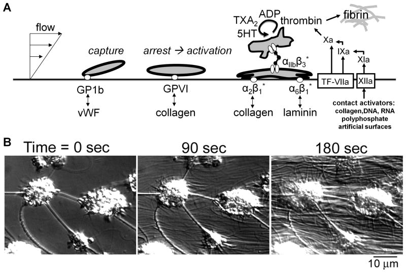
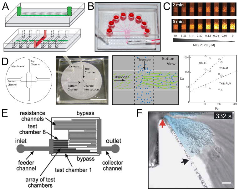
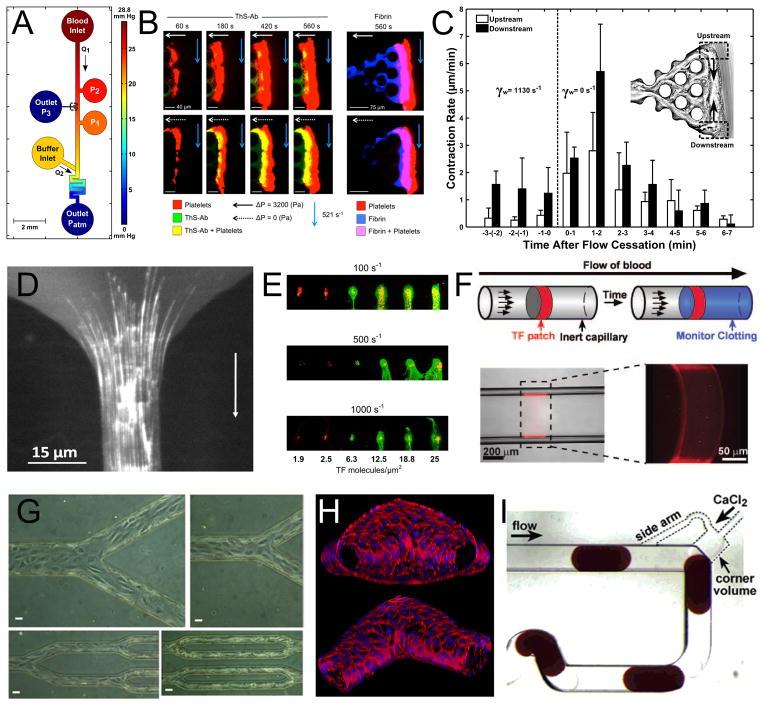
The study of blood ex vivo can occur in closed or open systems, with or without flow. Microfluidic devices, which constrain fluids to a small (typically submillimeter) scale, facilitate analysis of platelet function, coagulation biology, cellular biorheology, adhesion dynamics, and pharmacology and, as a result, can be an invaluable tool for clinical diagnostics. An experimental session can accommodate hundreds to thousands of unique clotting, or thrombotic, events. Using microfluidics, thrombotic events can be studied on defined surfaces of biopolymers, matrix proteins, and tissue factor, under constant flow rate or constant pressure drop conditions. Distinct shear rates can be generated on a device using a single perfusion pump. Microfluidics facilitated both the determination of intraluminal thrombus permeability and the discovery that platelet contractility can be activated by a sudden decrease in flow. Microfluidic devices are ideal for multicolor imaging of platelets, fibrin, and phosphatidylserine and provide a human blood analog to mouse injury models. Overall, microfluidic advances offer many opportunities for research, drug testing under relevant hemodynamic conditions, and clinical diagnostics.


Figures

Fig. 1
Fig. 1. Autocatalytic deposition of platelets on an injured vascular wall and generation of coagulation proteases
A, Adhesion of platelet to vWF mediates capture under arterial flow conditions, followed by platelet activation via GPVI. Once activated, the platelet integrins can bind collagen, laminin, and fibrinogen. Platelet activation is also associated with release of ADP and serotonin (5-HT), synthesis of thromboxane (TXA2) and exposure of phosphatidylserine which facilitates thrombin generation. Thrombin production is triggered primarily by tissue factor with contact activation via Factor XIIa having a secondary role in thrombosis. Thrombin also triggers the polymerization of fibrinogen to fibrin. B, Video microscopy of platelet aggregates forming on a surface with generation of fibrin strands.
Fig. 2
Fig. 2. Microfluidic devices for blood biology
A (top), A schematic drawing of a microfluidic channel casted in PDMS intended for patterning procoagulant proteins onto the surface of glass slides. (Bottom), The flow device, also in PDMS, is affixed onto the protein stripe and samples are perfused via syringe pump. Reproduced with permission from Reference . Copyright International Society on Thrombosis and Hemostasis. B, The 8-channel microfluidic device consists of 8 inlets perfused via syringe pump from one outlet. The device is adhered reversibly to a glass substrate via vacuum (bottom left tube) with a protein stripe patterned perpendicular to the region in which the 8 channels run parallel. Reproduced with permission from Reference . Copyright American Heart Association, Inc. C, Platelet adhesion is monitored in the 8-channel microfluidic device using epi-fluorescence microscopy. Here, fluorescently labeled platelets adhere to a collagen surface at venous shear rate in the presence of 8 different concentration of the P2Y1 antagonist, MRS 2179. [90] – Reproduced by permission of The Royal Society of Chemistry. D (left), A schematic representation of a 3 dimensional microfluidic device which allowed for the localized release of soluble agonists from one channel (bottom channel) to another channel (top channel) through a porous membrane. (Middle left), An image of this device. (Middle right), A drawing of the site of release of thrombin into a channel of flowing fibrinogen. The illustration indicates the generation of fibrin, which adhered to the membrane. (Right), In the thrombin release experiment it was found that the fibrin deposit formed had a morphology which was dependent on the ratios of reaction rate to convection (Da) and convection to diffusion (Pe). Reprinted from Reference with permission from the Biophysical Society. E, A schematic representation of a microfluidic device designed to study the role of shear stress in platelet adhesion. The image demonstrates that each of the test chambers is connected to a downstream channel of varying length which provides varying resistance and defines the flow rate (and therefore shear rate) in each of the chambers. Reproduced from Reference with permission of The Royal Society of Chemistry. F, In this microfluidic model a severe stenosis has been generated. Blood flow, from left to right, experiences extreme accelerating and decelerating flows as it traverses the stenosis apex (red arrow). As the platelets exit the high shear stress region they form a thrombus in the deceleration region (light blue). Reprinted by permission from Macmillan Publishers Ltd: Nature Biotechnology (Reference 99), copyright 2009.
Fig. 3
Fig. 3. Microfluidic Devices of Coagulation Research
A, A microfluidic model of bleeding designed for the study of thrombus permeability under flow. The device has integrated ports for localization of collagen and TF to the channel wall, pressure sensors, and buffer flow for precise pressure control. Blood flow (from top to bottom) will seep through the collagen scaffold connected to Outlet P3 until a platelet plug is formed. Reproduced with permission from Reference . Copyright American Heart Association, Inc. B, Epi-fluorescence microscopy performed at the collagen/TF scaffold of the device pictured in A reveals a large platelet aggregate (red) formed during perfusion which also stains positive for thrombin using a novel thrombin bio-sensor. Comparison of the top to bottom rows in which blood flow is allowed (top) or not allowed (bottom) to seep through Outlet P3 reveals that thrombin permeation into the clot is enhanced when it is not convected away. These clots also stain positive for fibrin (right). Reproduced with permission from Reference . Copyright International Society on Thrombosis and Hemostasis. C, Thrombus contraction was also observed in the microfluidic bleeding model. Here it is demonstrated that ~1 min after flow cessation the rate of clot retraction, measured by tracking the movement of trapped fluorescent beads, was 3-fold enhanced. Reproduced with permission from Reference . Copyright American Heart Association, Inc. D, A microfluidic model of stenosis reveals long fiber bundles of von Willebrand factor forming on a collagen type 1 surface under plasma flow at shear rates >30,000 s−1. Reproduced with permission from Reference . Copyright American Heart Association, Inc. E, Microcontact printing of collagen and varying amounts of tissue factor reveals a steep threshold for fibrin generation (green) under whole blood flow at varying shear rates in a parallel plate flow chamber construct. Platelets are labeled in red. This research was originally published in Reference . Copyright the American Society of Hematology. F, TF has also been patterned into microcapillary flow models using photolithographic techniques. The drawing illustrates that a soluble fluorescent reporter of thrombin will be activated downstream of the procoagulant patch. Reproduced with permission from Reference . Copyright Wolters Kluwer Health. G, The culture of confluent monolayers of endothelium has been achieved in microfluidic devices of PDMS (Republished with permission of the American Society for Clinical Investigation, from Reference 27) as well as on 3-dimensional collagen scaffolds (H). The in vitro vessels pictured in H were designed for studies involving permeability, angiogenesis, as well as thrombus formation after endothelial activation. Reproduced with permission from Reference . I, The microfluidic droplet reactor pictured here was designed to study clotting time in the presence of various inhibitors. Plugs of citrated whole blood enter at the top to which CaCl2 is added. The plug at the bottom left is entering the mixing region after which clotting will be assessed in a downstream region of the channel. Reprinted with permission from Reference . Copyright 2006 American Chemical Society.

Similar articles

Cited by

References

    1. Watson SP, Auger JM, McCarty OJ, Pearce AC. GPVI and integrin αIIbβ3 signaling in platelets. J Thromb Haemost. 2005;3:1752–62. - PubMed
    1. Ruggeri ZM, Mendolicchio GL. Adhesion mechanisms in platelet function. Circ Res. 2007;100:1673–85. - PubMed
    1. Flamm MH, Sinno T, Diamond SL. Simulation of aggregating particles in complex flows by the lattice kinetic Monte Carlo method. J Chem Phys. 2011;134:034905. - PMC - PubMed
    1. Laurenzi IJ, Diamond SL. Monte Carlo simulation of the heterotypic aggregation kinetics of platelets and neutrophils. Biophys J. 1999;77:1733–46. - PMC - PubMed
    1. Hockin MF, Jones KC, Everse SJ, Mann KG. A model for the stoichiometric regulation of blood coagulation. J Biol Chem. 2002;277:18322–33. - PubMed

Publication types