Review
. 2013:15:253-82.
doi: 10.1146/annurev-bioeng-071812-152409. Epub 2013 Apr 29.

Multifunctional nanoparticles for drug delivery and molecular imaging

Affiliations
Review

Multifunctional nanoparticles for drug delivery and molecular imaging

Gang Bao et al. Annu Rev Biomed Eng. 2013.

Abstract

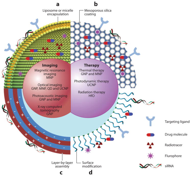
Recent advances in nanotechnology and growing needs in biomedical applications have driven the development of multifunctional nanoparticles. These nanoparticles, through nanocrystalline synthesis, advanced polymer processing, and coating and functionalization strategies, have the potential to integrate various functionalities, simultaneously providing (a) contrast for different imaging modalities, (b) targeted delivery of drug/gene, and (c) thermal therapies. Although still in its infancy, the field of multifunctional nanoparticles has shown great promise in emerging medical fields such as multimodal imaging, theranostics, and image-guided therapies. In this review, we summarize the techniques used in the synthesis of complex nanostructures, review the major forms of multifunctional nanoparticles that have emerged over the past few years, and provide a perceptual vision of this important field of nanomedicine.


Figures

Figure 1
Figure 1
Schematic diagram of multifunctional nanoparticles. Multifunctional nanoparticles can be generated by either combining nanocrystals with different functionalities or combining nanocrystals with functional small-molecule cargos through different surface engineering strategies. Four typical coatings developed for inorganic nanocrystals are (a) liposome or micelle encapsulation, (b) mesoporous silica coating, (c) layer-by-layer assembly, and (d ) surface conjugation. Abbreviations: GNP, gold nanoparticles; HfO, hafnium oxide nanoparticles; MNP, magnetic nanoparticles; QD, quantum dot; UCNP, upconversion nanoparticles.
Figure 2
Figure 2
Physical properties of inorganic nanoparticles. (a) Gold nanocage showing tunable surface plasmon resonance peak. (Reprinted from Reference with permission from Macmillan Publishers Ltd ©2007.) (b) Magnetic nanoparticles generated by doping iron oxide with various magnetic ions. These nanoparticles exhibit different mass magnetization and, subsequently, MRI T2 relaxivity. (Reprinted from Reference with permission from Macmillan Publishers Ltd © 2007.) (c) Quantum dots showing size-tunable fluorescence emission. (Reprinted from Reference with permission from Macmillan Publishers Ltd © 2005.) (d ) Emission spectra of upconversion nanoparticles with different sensitizer and activator ions. (Reprinted with permission from Reference , © 2008 American Chemical Society.) Abbreviations: λl, wavelength; CLIO, cross-linked iron oxide; MEIO, magnetism-engineered iron oxide; MRI, magnetic resonance imaging; TEM, transmission electron microscopy; ABS, absorption spectrum; EM, emission.
Figure 3
Figure 3
Trimodality nanoparticle for pre- and intraoperative brain tumor imaging. (a) Trimodality detection of brain tumors in living mice. (b) Raman-guided intraoperative surgery. Abbreviations: CD11b, cluster of differentiation molecule 11b; GFP, green fluorescent protein; MRI, magnetic resonance imaging. (Reprinted from Reference with permission from Macmillan Publishers Ltd © 2012.)
Figure 4
Figure 4
Image-guided siRNA delivery. (a) Schematic diagram of the siRNA carrier with MRI/NIR modalities (MN-NIRF-siRNA). MN core and Cy5.5 can be detected with MRI and NIRF imaging, respectively. (b) In vivo MRI of mice bearing subcutaneous LS174T human colorectal adenocarcinoma (arrows). The significant drop in T2 relaxivity after administration of the contrast agent (P = 0.003) confirmed probe delivery. (c) NIRF imaging of the mouse following injection of MN-NIRF-siSurvivin (top: white light, middle: NIRF, bottom: color-coded overlay). (d ) The tumor treated with MN-NIRF-siSurvivin (left) showed distinct areas with a high density of apoptotic nuclei ( green). Sections were counterstained with DAPI. (e) H&E staining of frozen tumor sections revealed considerable eosinophilic areas of tumor necrosis (N) in tumors treated with MN-NIRF-siSurvivin (left). Purple hematoxiphilic regions (V) indicate viable tumor tissues. (f) Quantitative RT-PCR analysis of survivin expression in LS174T tumors after injection with either MN-NIRF-siSurvivin, a mismatch control, or the parental MN. Abbreviations: DAPI, diamidino-2-phenylindole; GFP, green fluorescent protein; H&E, ematoxylin and eosin; MN, magnetic nanoparticle; MPAP, myristoylated polyarginine peptide; NIRF, near-infrared fluorescence; RT-PCR, real-time polymerase chain reaction. (Reprinted from Reference with permission from Macmillan Publishers Ltd © 2007.)
Figure 5
Figure 5
Magnetic targeting of lentiviral vectors and positioning of transduced cells. (a) The magnetic flux density of magnets placed next to a vessel. (b) Magnetic targeting of lentiviral vectors to aorta during ex vivo perfusion. (c) In vivo positioning of lentivirus/magnetic nanoparticle–transduced HUVECs (human umbilical vein endothelial cells) to the intimae of injured common carotid arteries by magnetic forces. (Reprinted with permission from Reference , © 2009 National Academy of Sciences.)
Figure 6
Figure 6
Magnetically activated release system (MARS). (a) Schematic diagram of nanoparticles, machines, and assembly. Magnetic-core silica nanoparticles (MCSN) are synthesized by coating zinc-doped iron oxide nanocrystals (1) with mesoporous silica (2). The base of the molecular machine is then attached to the nanoparticle surface (3). The drug is loaded into the particle and capped (4) to complete the system. Release can be realized using remote heating via the introduction of an oscillating magnetic field (5). (b) Fluorescent microscope images (1, 3, and 5) and fluorescent images overlaid with differential interference contrast (DIC) (2, 4, and 6). Images 5 and 6 demonstrate doxorubicin (DOX) release after a 5-min AC field exposure. Color scheme: green, fluorescently labeled MARS; red, DOX; yellow, merged green and red. (c) Quantification of cell death after treatments shown in Panel b. (Reprinted with permission from Reference , © 2010 American Chemical Society.)

References

    1. Davis ME, Chen ZG, Shin DM. Nanoparticle therapeutics: an emerging treatment modality for cancer. Nat Rev Drug Discov. 2008;7:771–82. - PubMed
    1. Peer D, Karp JM, Hong S, Farokhzad OC, Margalit R, Langer R. Nanocarriers as an emerging platform for cancer therapy. Nat Nanotechnol. 2007;2:751–60. - PubMed
    1. Kim BY, Rutka JT, Chan WC. Nanomedicine. N Engl J Med. 2010;363:2434–43. - PubMed
    1. Hrkach J, Von Hoff D, Mukkaram Ali M, Andrianova E, Auer J, et al. Preclinical development and clinical translation of a PSMA-targeted docetaxel nanoparticle with a differentiated pharmacological profile. Sci Transl Med. 2012;4:128ra39. - PubMed
    1. Gratton SE, Williams SS, Napier ME, Pohlhaus PD, Zhou Z, et al. The pursuit of a scalable nanofabrication platform for use in material and life science applications. Acc Chem Res. 2008;41:1685–95. - PMC - PubMed

Publication types