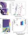
Functional attachment of soft tissues to bone: development, healing, and tissue engineering
- PMID: 23642244
- PMCID: PMC3925419
- DOI: 10.1146/annurev-bioeng-071910-124656
Functional attachment of soft tissues to bone: development, healing, and tissue engineering
Erratum in
- Annu Rev Biomed Eng. 2013;15:vi
Abstract
Connective tissues such as tendons or ligaments attach to bone across a multitissue interface with spatial gradients in composition, structure, and mechanical properties. These gradients minimize stress concentrations and mediate load transfer between the soft and hard tissues. Given the high incidence of tendon and ligament injuries and the lack of integrative solutions for their repair, interface regeneration remains a significant clinical challenge. This review begins with a description of the developmental processes and the resultant structure-function relationships that translate into the functional grading necessary for stress transfer between soft tissue and bone. It then discusses the interface healing response, with a focus on the influence of mechanical loading and the role of cell-cell interactions. The review continues with a description of current efforts in interface tissue engineering, highlighting key strategies for the regeneration of the soft tissue-to-bone interface, and concludes with a summary of challenges and future directions.
Figures


References
-
- Woo SL, Maynard J, Butler DL, Lyon RM, Torzilli PA, et al. Ligament, tendon, and joint capsule insertions to bone. In: Woo SL, Bulkwater JA, editors. Injury and Repair of the Musculoskeletal Soft Tissues. Rosemont, IL: Am. Acad. Orthop. Surg; 1988. pp. 133–66.
-
- Thomopoulos S, Williams GR, Gimbel JA, Favata M, Soslowsky LJ. Variations of biomechanical, structural, and compositional properties along the tendon to bone insertion site. J Orthop Res. 2003;21:413–19. - PubMed
-
- Thomopoulos S, Marquez JP, Weinberger B, Birman V, Genin GM. Collagen fiber orientation at the tendon to bone insertion and its influence on stress concentrations. J Biomech. 2006;39:1842–51. - PubMed
-
- Lu HH, Jiang J. Interface tissue engineering and the formulation of multiple-tissue systems. Adv Biochem Eng Biotechnol. 2006;102:91–111. - PubMed
Publication types
MeSH terms
Substances
Grants and funding
- U01 EB016422/EB/NIBIB NIH HHS/United States
- R01 AR055580/AR/NIAMS NIH HHS/United States
- R21-AR056459/AR/NIAMS NIH HHS/United States
- R21 AR052402/AR/NIAMS NIH HHS/United States
- R01 AR060820/AR/NIAMS NIH HHS/United States
- R01-AR057836/AR/NIAMS NIH HHS/United States
- R21 AR056459/AR/NIAMS NIH HHS/United States
- R01-AR055580/AR/NIAMS NIH HHS/United States
- R01 AR057836/AR/NIAMS NIH HHS/United States
- R01 AR055280/AR/NIAMS NIH HHS/United States
- R01-AR060820/AR/NIAMS NIH HHS/United States
- R21-AR052402/AR/NIAMS NIH HHS/United States
LinkOut - more resources
Full Text Sources
Other Literature Sources

