. 2013 May 4;20(1):27.
doi: 10.1186/1423-0127-20-27.

Proteomic analyses and identification of arginine methylated proteins differentially recognized by autosera from anti-Sm positive SLE patients

Affiliations

Proteomic analyses and identification of arginine methylated proteins differentially recognized by autosera from anti-Sm positive SLE patients

Hong-How Chang et al. J Biomed Sci. .

Abstract

Background: Antibodies against spliceosome Sm proteins (anti-Sm autoantibodies) are specific to the autoimmune disease systemic lupus erythematosus (SLE). Anti-Sm autosera have been reported to specifically recognize Sm D1 and D3 with symmetric di-methylarginines (sDMA). We investigated if anti-Sm sera from local SLE patients can differentially recognize Sm proteins or any other proteins due to their methylation states.

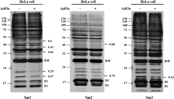
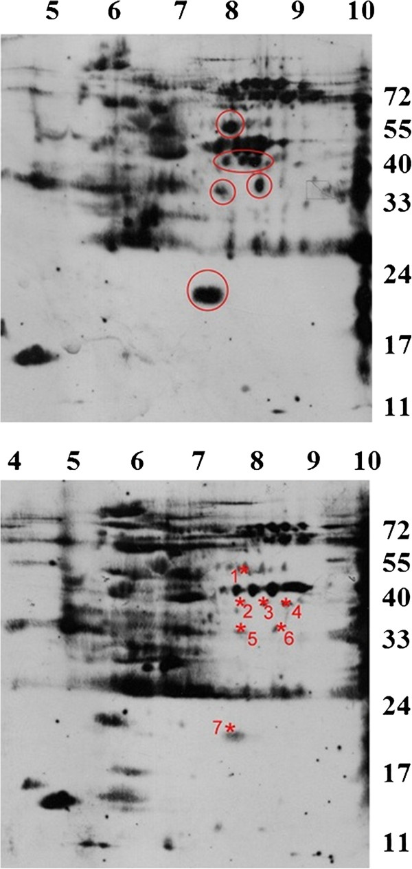
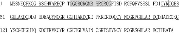
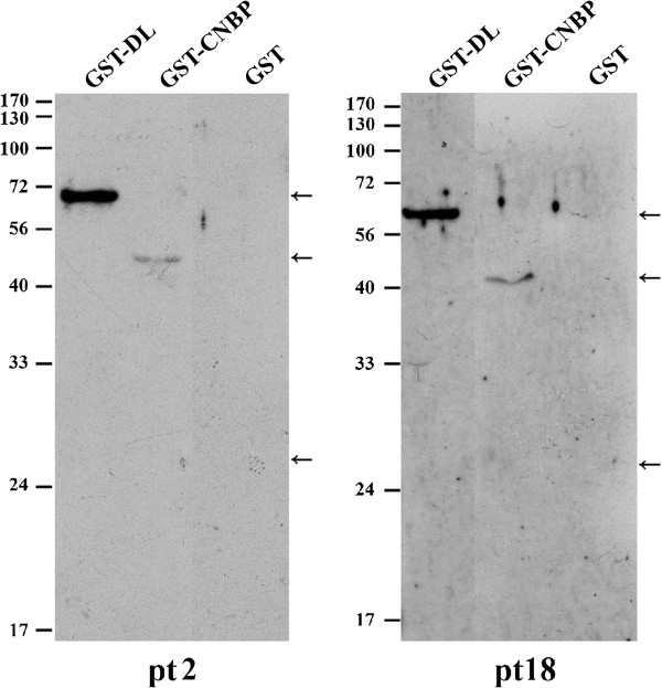
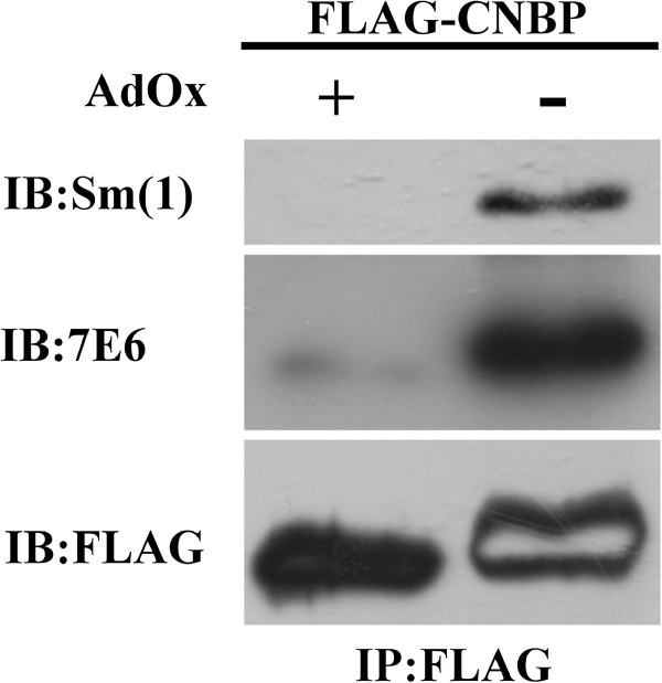
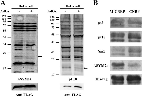
Results: We prepared HeLa cell proteins at normal or hypomethylation states (treated with an indirect methyltransferase inhibitor adenosine dialdehyde, AdOx). A few signals detected by the anti-Sm positive sera from typical SLE patients decreased consistently in the immunoblots of hypomethylated cell extracts. The differentially detected signals by one serum (Sm1) were pinpointed by two-dimensional electrophoresis and identified by mass spectrometry. Three identified proteins: splicing factor, proline- and glutamine-rich (SFPQ), heterogeneous nuclear ribonucleoprotein D-like (hnRNP DL) and cellular nucleic acid binding protein (CNBP) are known to contain methylarginines in their glycine and arginine rich (GAR) sequences. We showed that recombinant hnRNP DL and CNBP expressed in Escherichia coli can be detected by all anti-Sm positive sera we tested. As CNBP appeared to be differentially detected by the SLE sera in the pilot study, differential recognition of arginine methylated CNBP protein by the anti-Sm positive sera were further examined. Hypomethylated FLAG-CNBP protein immunopurified from AdOx-treated HeLa cells was less recognized by Sm1 compared to the CNBP protein expressed in untreated cells. Two of 20 other anti-Sm positive sera specifically differentiated the FLAG-CNBP protein expressed in HeLa cells due to the methylation. We also observed deferential recognition of methylated recombinant CNBP proteins expressed from E. coli by some of the autosera.

Conclusion: Our study showed that hnRNP DL and CNBP are novel antigens for SLE patients and the recognition of CNBP might be differentiated dependent on the level of arginine methylation.


Figures

Figure 1
Figure 1
Differential recognition of proteins due to methylation status by anti-Sm autosera. HeLa cell extracts (30 μg of total protein) were prepared from cells grown in the presence or absence of 20 μM of AdOx. The cell extracts were separated by SDS-PAGE and transferred onto nitrocellulose membrane. Western blot analysis was performed using Sm1, Sm2 and Sm3 autosera. The positions of the differentially detected signals are indicated by arrows and the intensity ratios are shown. The positions of typical anti-Sm recognized SmB/B′, SmD1 and D3 proteins are also indicated.
Figure 2
Figure 2
Resolving differentially recognized proteins by Sm1 autosera due to methylation status by 2-DE. HeLa cell extracts (250 μg of total protein) treated with AdOx (lower panel) or not (upper panel) were resolved by 2-DE, blotted and detected by anti-Sm1. The other parallel gel was stained by SyproRuby. The differentially detected signals that can match to protein spots are indicated by red asteroid and numbered.
Figure 3
Figure 3
The amino acid sequence of human CNBP. The seven Cys-Cys-His-Cys (CCHC) type zinc knuckle domains (C-Φ-X-C-G-X3-H-X4-C, where Φis an aromatic amino acid and X is a variable amino acid) in the human CNBP protein are underlined. The RG sequence between zinc finger ZF1 and ZF2 is indicated with shading. The peptide sequence identified by MS/MS is boxed.
Figure 4
Figure 4
Recognition of hnRNP DL and CNBP by anti-Sm positive SLE patient sera. GST-fused hnRNP DL and CNBP were prepared as described in Methods. The GST-fused proteins or GST were applied to SDS-PAGE and the immunoblots detected by the autosera from patient (pt) 2 and 18 are shown.
Figure 5
Figure 5
CNBP is methylated in vivo and is recognized differentially by Sm1 due to its methylation status. HeLa cells were transfected with pFLAG-CNBP plasmids. The FLAG-tagged proteins were immunoprecipitated by anti-FLAG agarose. Proteins eluted by the FLAG peptide were then analyzed by Western blot analyses with anti-Sm1. The blot was stripped of the interacted antibodies and then probed with a mono- and di-methylarginine-specific antibody 7E6. The same blot was stripped and then re-probed with the anti-FLAG antibody. The internal blank region of the FLAG-CNBP signal of the AdOx- sample detected by anti-FLAG was due to previous stripping.
Figure 6
Figure 6
Differential detection of CNBP protein by anti-Sm positive SLE sera. (A) HeLa cell extracts prepared from cells that were transfected with the pFLAG-CNBP plasmid and treated with AdOx or not were analyzed by Western blot analyses with an aDMA-specific antibody ASYM24 (left panel) or autosera from SLE patient 18 (right panel) and re-probed with anti-FLAG antibodies. The positions of FLAG-CNBP are indicated by arrows. (B) (His)6-CNBP proteins prepared from E. coli cells co-expressing PRMT1(M-CNBP) or not (CNBP) were analyzed by Western blot analyses. Differential detection of (His)6-CNBP by autosera from patient 5, 18 and Sm1 are shown. Arginine methylation of CNBP was confirmed by the detection with ASYM24. The blot was stripped and re-probed with anti-(His)6 (His-tag) antibodies.

Similar articles

Cited by

References

    1. Atassi MZ, Casali P. Molecular mechanisms of autoimmunity. Autoimmunity. 2008;41:123–132. doi: 10.1080/08916930801929021. - DOI - PubMed
    1. Anderton SM. Post-translational modifications of self antigens: implications for autoimmunity. Curr Opin Immunol. 2004;16:753–758. doi: 10.1016/j.coi.2004.09.001. - DOI - PubMed
    1. Doyle HA, Mamula MJ. Posttranslational modifications of self-antigens. Ann N Y Acad Sci. 2005;1050:1–9. doi: 10.1196/annals.1313.001. - DOI - PubMed
    1. Doyle HA, Mamula MJ. Autoantigenesis: the evolution of protein modifications in autoimmune disease. Curr Opin Immunol. 2012;24:112–118. doi: 10.1016/j.coi.2011.12.003. - DOI - PMC - PubMed
    1. Bedford MT, Clarke SG. Protein arginine methylation in mammals: who, what, and Why. Mol Cell. 2009;33:1–13. doi: 10.1016/j.molcel.2008.12.013. - DOI - PMC - PubMed

Publication types

MeSH terms