. 2013 May 4:11:111.
doi: 10.1186/1479-5876-11-111.

SATB1 is overexpressed in metastatic prostate cancer and promotes prostate cancer cell growth and invasion

Affiliations

SATB1 is overexpressed in metastatic prostate cancer and promotes prostate cancer cell growth and invasion

Lijun Mao et al. J Transl Med. .

Abstract

Background: Special AT-rich sequence binding protein 1 (SATB1) is a nuclear factor that functions as the global chromatin organizer to regulate chromatin structure and gene expression gene expression. SATB1 has been shown to be abnormally expressed in various types of cancer. However, the expression and role of SATB1 in prostate cancer remain unclear.

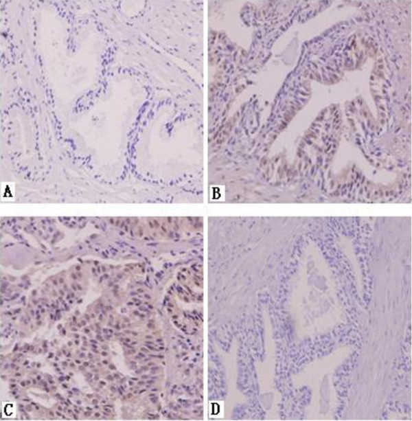
Methods: 120 cases of prostatic carcinoma and 60 cases of benign prostate hyperplasia were analyzed for SATB1 expression by immunohistochemistry. LNCaP, DU-145, and PC3 prostate cancer cells were examined for SATB1 expression by Western blot analysis. Cell proliferation and invasion was evaluated by CCK8 and transwell invasion assay, respectively.

Results: SATB1 staining was stronger in prostatic carcinomas with metastasis than in those without metastasis, but was absent in benign prostate hyperplasia. Furthermore, SATB1 expression was positively correlated with bone metastasis and the Gleason score. SATB1 overexpression promoted the proliferation and invasion of LNCaP cells while SATB1 knockdown inhibited the proliferation and invasion of DU-145 cells.

Conclusions: These findings provide novel insight into oncogenic role of SATB1 in prostate cancer, suggesting that SATB1 is a promising biomarker and therapeutic target for prostate cancer.


Figures

Figure 1
Figure 1
SATB1 immunohistochemistry staining. A, benign prostate hyperplasia. B, prostatic carcinoma with metastasis. C, prostatic carcinoma without metastasis. D, negative control. Magnification: 400 × .
Figure 2
Figure 2
SATB1 expression is correlated with the invasion ability of prostate cancer cells. A-C: The invasion of LNCaP, DU-145 and PC-3 cells was measured by Transwell chamber. A, DU-145. B, PC-3.C, LNCaP. Magnification: 200×. D: Western blot analysis of the relative protein level of SATB1 in LNCaP, DU-145 and PC-3 cells (n = 3). β-actin served as loading control. *P < 0.05 compared to DU-145 cells. Note that SATB1 antibody could not detect SATB2 protein in the cell lysates.
Figure 3
Figure 3
Silencing of SATB1 in DU-145 cells. DU-145 cells were untransfected (control), transfected with pSliencer3.1 control vector or transfected with pSilencer3.1-SATB1 vector for 24 h, 48 h, or 72 h. The protein levels of SATB1 and SATB2 were detected by Western blot analysis (n = 3). β-actin served as loading control. *P < 0.05 compared to cells transfected with pSliencer3.1.
Figure 4
Figure 4
Silencing of SATB1 inhibits the invasion and growth of DU-145 cells. A-D: The invasion of DU-145 cells was measured by Transwell chamber. A, DU-145 cells untransfected. B, DU-145 cells transfected with pSliencer3.1. C, DU-145 cells transfected with pSilencer3.1-SATB1. Magnification: 200×. D: Quantitative analysis of the number of invaded cells as shown in A-C (n = 3). E: The proliferation of DU-145 cells was determined by CCK8 assay (n = 3). The cell proliferation inhibition rate was calculated for DU-145 cells at 24 h, 48 h and 72 h after transfection with pSilencer3.1-SATB1. *P < 0.05 compared to cells transfected with pSliencer3.1.
Figure 5
Figure 5
Reconstitution of SATB1 expression in LNCaP cells. LNCaP cells were untransfected (control), transfected with pcDNA3.1 control vector or transfected with pcDNA3.1-SATB1 vector for 12 h, 24 h, or 36 h. A. The mRNA level of SATB1 was detected by RT-PCR (n = 3). B. The protein level of SATB1 was detected by Western blot analysis (n = 3). β-actin served as loading control. *P < 0.05 compared to cells transfected with pcDNA3.1.
Figure 6
Figure 6
Overexpression of SATB1 promotes the invasion and growth of LNCaP cells. A-D: The invasion of LNCaP cells was measured by Transwell chamber. A, LNCaP cells untransfected. B, LNCaP cells transfected with pcDNA3.1. C, LNCaP cells transfected with pcDNA3.1-SATB1. Magnification: 200×. D: Quantitative analysis of the number of invaded cells as shown in A-C (n = 3). *P < 0.05 compared to cells transfected with pcDNA3.1. E: The proliferation of LNCaP cells was determined by CCK8 assay (n = 3). The cell proliferation ratio was calculated for LNCaP cells at 24 h, 48 h and 72 h after transfection of pcDNA3.1 or pcDNA3.1-SATB1. *P < 0.05 compared to corresponding cells transfected with pcDNA3.1.

Similar articles

Cited by

References

    1. Saad F, Pantel K. The current role of circulating tumor cells in the diagnosis and management of bone metastases in advanced prostate cancer. Future Oncol. 2012;8:321–331. doi: 10.2217/fon.12.3. - DOI - PubMed
    1. Raheem O, Kulidjian AA, Wu C, Jeong YB, Yamaguchi T, Smith KM, Goff D, Leu H, Morris SR, Cacalano NA, Masuda K, Jamieson CH, Kane CJ, Jamieson CA. A novel patient-derived intra-femoral xenograft model of bone metastatic prostate cancer that recapitulates mixed osteolytic and osteoblastic lesions. J Transl Med. 2011;9:185. doi: 10.1186/1479-5876-9-185. - DOI - PMC - PubMed
    1. Galande S, Purbey PK, Notani D, Kumar PP. The third dimension of gene regulation: organization of dynamic chromatin loopscape by SATB1. Curr Opin Genet Dev. 2007;17:408–414. doi: 10.1016/j.gde.2007.08.003. - DOI - PubMed
    1. Chu SH, Ma YB, Feng DF, Zhang H, Zhu ZA, Li ZQ, Jiang PC. Upregulation of SATB1 is associated with the development and progression of glioma. J Transl Med. 2012;10:149. doi: 10.1186/1479-5876-10-149. - DOI - PMC - PubMed
    1. Meng WJ, Yan H, Zhou B, Zhang W, Kong XH, Wang R, Zhan L, Li Y, Zhou ZG, Sun XF. Correlation of SATB1 overexpression with the progression of human rectal cancer. Int J Colorectal Dis. 2012;27:143–150. doi: 10.1007/s00384-011-1302-9. - DOI - PubMed

Publication types

MeSH terms