. 2013 May 2;12(5):559-72.
doi: 10.1016/j.stem.2013.04.008.

Directed differentiation and functional maturation of cortical interneurons from human embryonic stem cells

Affiliations

Directed differentiation and functional maturation of cortical interneurons from human embryonic stem cells

Asif M Maroof et al. Cell Stem Cell. .

Abstract

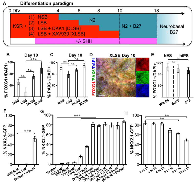
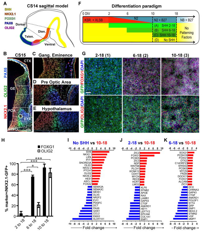
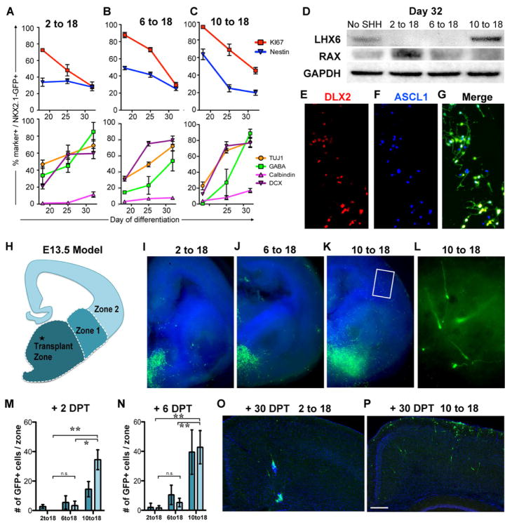
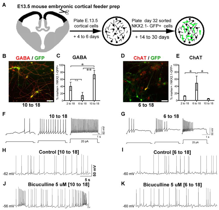
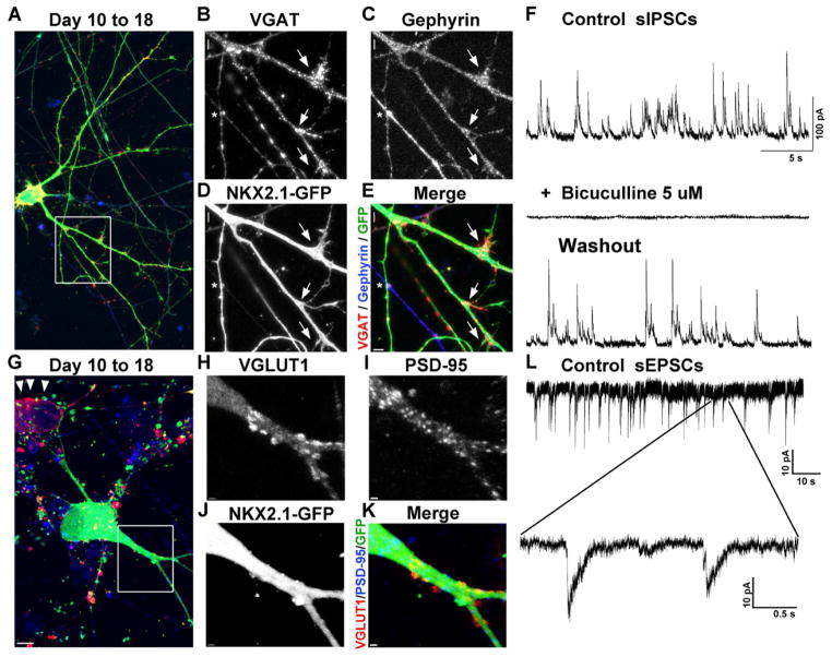
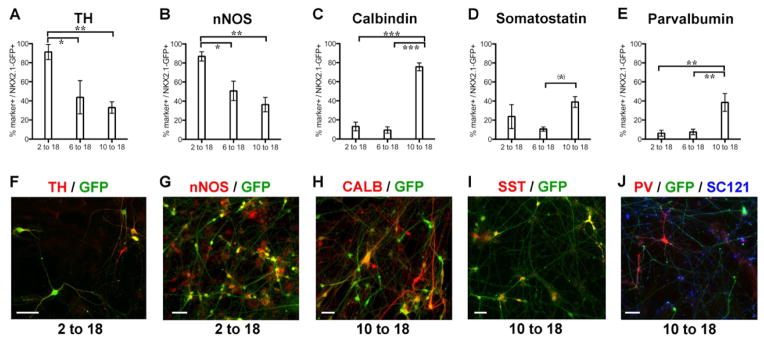
Human pluripotent stem cells are a powerful tool for modeling brain development and disease. The human cortex is composed of two major neuronal populations: projection neurons and local interneurons. Cortical interneurons comprise a diverse class of cell types expressing the neurotransmitter GABA. Dysfunction of cortical interneurons has been implicated in neuropsychiatric diseases, including schizophrenia, autism, and epilepsy. Here, we demonstrate the highly efficient derivation of human cortical interneurons in an NKX2.1::GFP human embryonic stem cell reporter line. Manipulating the timing of SHH activation yields three distinct GFP+ populations with specific transcriptional profiles, neurotransmitter phenotypes, and migratory behaviors. Further differentiation in a murine cortical environment yields parvalbumin- and somatostatin-expressing neurons that exhibit synaptic inputs and electrophysiological properties of cortical interneurons. Our study defines the signals sufficient for modeling human ventral forebrain development in vitro and lays the foundation for studying cortical interneuron involvement in human disease pathology.


Figures

Figure 1
Figure 1. Wnt inhibition and activation of SHH signaling yields highly efficient derivation of forebrain fates and NKX2.1 induction
(A) Schematic of the differentiation protocol in the dual SMAD inhibition paradigm to generate anterior neural progenitors. NSB: Noggin+ SB431542. LSB: LDN193189+ SB31542. B–E): When either DKK1 or XAV939, both Wnt signaling antagonists, were added to the dual SMAD inhibition protocol (DLSB or XLSB), there was a significant increase in the percentage of FOXG1+ cells (B) without loss of PAX6 expression (C): ** p < 0.01; *** p < 0.001; using ANOVA followed by Scheffe test. D) Representative immunofluorescent image for FOXG1 (red) and PAX6 (green) expression at day 10 following XLSB treatment. Single channel fluorescent images of marked region are shown in the three right panels E) Robust telencephalic specification using XLSB was also observed at comparable efficiencies in human induced pluripotent stem cells (hiPSC lines SeV6, C72; n = 4). F–H) Addition of SHH signaling to the XLSB protocol significantly enhanced the production of NKX2.1::GFP expressing progenitors. (F) 5nM SHH (Sonic C24II) and 1μm Purmorphamine, added from day 4, showed synergistic effects in inducing NKX2.1::GFP expression at day 10 (*** p < 0.001; compared to SHH). A range of concentrations of SHH and Purmorphamine are compared at day 18 in (G), and again co-treatment was greatly superior to quite high concentrations of either SHH or purmorphamine alone (*** p < 0.001; compared to no SHH using ANOVA followed by Scheffe test). (H) Delaying the timing of SHH exposure between 2 and 10 days of differentiation did not dramatically affect the efficiency of NKX2.1::GFP induction measured at day 18 (*** p < 0.001 compared to 0–18 using ANOVA followed by Scheffe test). P: purmorphamine, S: Sonic hedgehog. Data are from hESC line HES-3 (NKX2.1::GFP) in panels B,C,F,G,H from hESC line WA-09/H9 (panel D) and from hESC line WA-09/H9 and hiPSC lines SeV6 and C72 (panel E). Scale bar in (D) represents 125μm. Data in (B,C,E–H) are represented as mean ± SEM.
Figure 2
Figure 2. Timing of SHH exposure determines the regional identity of NKX2.1::GFP expressing progenitors
A) Model of human prosencephalon (sagittal view at Carnegie stage 14 (CS14)) with expression of forebrain patterning markers based on published data (Kerwin et al., 2010). B–E) Coronal (oblique) hemisection of the human prosencephalon at Carnegie stage 15 (CS15) demonstrate expression of NKX2.1, OLIG2, and PAX6. NKX2.1 and OLIG2 are expressed in various regions throughout the ventral prosencephalon, whereas PAX6 is restricted to the dorsal prosencephalon and the eye. The expression of these proteins is non-overlapping, except in the ganglionic eminence (C) where OLIG2 and NKX2.1 are co-expressed. The scale bar in (B) represents 200μm. F) Schematic illustration of the distinct time periods of SHH and purmorphamine treatment used in combination with the XLSB protocol. G,H) Immunofluorescence for OLIG2 and FOXG1 in NKX2.1::GFP line at day 18 of differentiation. Treatment with SHH after day 6 (6-18 and 10-18 group) significantly increases the percentage of NKX2.1::GFP+ cells that co-express FOXG1 as quantified in (H). The scale bar in (G) represents 50μm Treatment with SHH after day 10 (10-18 group) enhanced the derivation of NKX2.1::GFP+ cells co-expressing OLIG2, (data are mean ± SEM; * p < 0.05, *** p < 0.001, compared to 2-18 using ANOVA followed by Scheffe test). Expression of FOXG1, NKX2.1 and OLIG2 indicates a pattern characteristic of ganglionic eminence (Tekki-Kessaris et al.). I–K) Microarray data from cells sorted for NKX2.1::GFP expression at day 18 of differentiation, comparing gene expression levels between the SHH day 10-18 protocol versus no SHH (I), day 10-18 versus day 2-18 protocol (J), and day 10-18 versus 6-18 protocol (K). Red bars indicate genes expressed at higher levels in the SHH 10-18 protocol, blue bars indicate genes expressed at lower levels in day 10-18 protocol. All changes are significant at p < 0.001. Figure 2, see also Figure S1 & Table S1.
Figure 3
Figure 3. Conversion from cycling neural progenitors to neuronal precursors and the assessment of their migratory potential
A–C) At day 18 cells from the three indicated protocols were subjected to FACS for NKX2.1::GFP expression then replated and evaluated for co-labeling with markers indicated. For all three protocols there was a decline in co-labeling with markers of progenitors (upper panels: red line, nestin; blue line Ki67), and an increase in markers of neuronal differentiation (lower panels: green, GABA; yellow, TUJ1; purple, doublecortin (DCX); pink, calbindin). Data are mean ± SEM. D) Western blotting showed an increase in the hypothalamic-enriched protein RAX in the 2 to 18 condition, and an increase in the medial ganglionic eminence (MGE)-enriched protein LHX6 in the 10 to 18 condition. Cells were sorted for NKX2.1::GFP prior to analysis. E–G) At day 32, many of the NKX2.1::GFP+ cells from the 10 to 18 condition also expressed DLX2 and ASCL1. H–N) Grafting of day 32-sorted NKX2.1::GFP+ cells into the MGE of E13.5 coronal mouse slice cultures. H) Schematic of coronal hemisection demonstrating the site of transplantation and the zones for quantification of migration. I,J) In both the 2 to 18 and the 6 to 18 conditions, very few cells migrated into zone 1 and fewer into zone 2. K,L) Only the 10 to 18 condition demonstrated significant and robust migration into the cortical and striatal regions, with many GFP+ cells exhibiting bipolar morphologies consistent with a migratory cell (L). The regions where GFP+ cells were detected were quantified two (M) and six (N) days post transplantation DPT (data are mean ± SEM; * p < 0.05; ** p < 0.01 using ANOVA followed by Scheffe test). O,P) Transplantation of day 32 sorted NKX2.1::GFP+ cells (day 10 to 18 protocol) into the neocortex of neonatal mice followed by their evaluation in fixed sections at postnatal day 30. In marked contrast to the MGE-like cells from the SHH 10-18 protocol (P), neither the SHH 2-18 protocol (O) nor the SHH 6-18 protocol (not shown) resulted in extensive migration from the graft site. The scale bar in (P) represents 200μm. See also Figure S2 & Figure S3
Figure 4
Figure 4. Maturation of NKX2.1+ cells into physiologically active neurons
A) Preparation of cortical excitatory neuron cultures from embryonic day 13.5 (E13.5) mice, onto which human NKX2.1::GFP+ cells (after FACS at day 32) are plated. The co-culture system was critical for promoting neuronal maturation given the protracted in vivo maturation rates of NKX2.1+ cells. B–E) After 30 days in vitro (DIV), cultures from the SHH 10-18 conditions are enriched for NKX2.1::GFP+ cells that co-express GABA (B, quantified in C: mean ± SEM; * p < 0.05; ** p < 0.01 using ANOVA followed by Scheffe test). In contrast only the SHH 6-18 condition was enriched for NKX2.1::GFP co-labeling with choline acetyl transferase (ChAT) (D, quantified in E: mean ± SEM; * p < 0.05 using ANOVA followed by Scheffe test). F, G) Spiking patterns of SHH day 10-18 (F) and SHH day 6-18 (G) neurons recorded at 28 DIV. Action potentials were initiated by protocols shown at bottom. H–K) Spontaneous spiking was recorded from cultures enriched for GABAergic (SHH day 10 to 18) and cholinergic (6 to 18) neurons in the absence (H, I) and the presence (J,K) of the GABAA receptor antagonist bicuculline. Bicuculline had little effect on the spontaneous firing activity in the 6 to 18 condition, consistent with the lack of GABAergic cells from either the mouse feeder or the human NKX2.1::GFP+ cells generated by this protocol. The scale bars (BD) represent 50μm.
Figure 5
Figure 5. NKX2.1::GFP+ GABAergic interneurons receive both excitatory and inhibitory synaptic inputs
A–E) Collapsed z-stack confocal image showing NKX2.1::GFP+, vesicular GABA transporter (VGAT; red in A), and the post-synaptic GABAergic marker gephyrin (blue in A). The dendrites of this GFP+ cell that co-label with gephyrin are receiving VGAT-expressing pre-synaptic terminals (arrows). In addition, a GFP+ axonal process formed a VGAT+ pre-synaptic terminal adjacent to a GFP negative, gephyrin-expressing post-synaptic process (asterisk). F) Whole-cell patch clamp reveals spontaneous inhibitory postsynaptic currents (sIPSCs) recorded from an NKX2.1::GFP+ neuron (SHH 10 to 18 protocol), which are reversibly blocked by the addition of the GABA-A receptor antagonist bicuculline. G-K) Collapsed z-stack confocal image showing NKX2.1::GFP, vesicular glutamate transporter 1 (VGLUT1; red in G), and the post-synaptic marker PSD-95 (blue; C). This GFP+ cell has dendrites that co-label with PSD-95 that are adjacent to VGLUT1-expressing pre-synaptic terminals. Note the presence of a GFP negative cell expressing VGLUT1 (red; G arrowheads), confirming the presence of excitatory glutamatergic neurons in the culture. L) Consistent with the apparent presence of glutamatergic synaptic inputs, spontaneous excitatory postsynaptic currents (sEPSCs) were detected in the NKX2.1::GFP+ neurons (10 to 18). All cells were plated on mouse cortical feeder following FACS for NKX2.1::GFP at day 32. See also Figure S4.
Figure 6
Figure 6. Neurochemical profiling of NKX2.1::GFP+ cells grown on mouse cortical feeders for 30 DIV
Cells were labeled by immunofluorescence for the markers indicated, and results quantified in graphs (A–E: mean ± SEM; * p < 0.05, ** p < 0.01, *** p < 0.001 using ANOVA followed by Scheffe test: (*) p < 0.05 when compared directly to 6-18 group. p-value did not reach significance in standard Scheffe test: p = 0.08). Panels (F–J) show representative images of cellular labeling. In the SHH 2 to 18 condition, most of the cells co-labeled with tyrosine hydroxylase (TH; A, F) and nNOS (B, G). In the 10 to 18 condition, many of the GFP+ cells co-labeled with calbindin (Calb; C, H), somatostatin (SST; D, I), and parvalbumin (E, J) each of which is present in subpopulations of mature cortical interneurons in humans. All cells were plated on mouse cortical feeder following FACS for NKX2.1::GFP at day 32. The scale bars (F–J) represent 50μm. See also Figure S5, Figure S6 & Table S2.

Comment in

References

    1. Anderson SA, Classey JD, Conde F, Lund JS, Lewis DA. Synchronous development of pyramidal neuron dendritic spines and parvalbumin-immunoreactive chandelier neuron axon terminals in layer III of monkey prefrontal cortex. Neuroscience. 1995;67:7–22. - PubMed
    1. Anderson SA, Eisenstat DD, Shi L, Rubenstein JL. Interneuron migration from basal forebrain to neocortex: dependence on Dlx genes. Science. 1997;278:474–476. - PubMed
    1. Anderson SA, Marin O, Horn C, Jennings K, Rubenstein JL. Distinct cortical migrations from the medial and lateral ganglionic eminences. Development. 2001;128:353–363. - PubMed
    1. Baraban SC, Southwell DG, Estrada RC, Jones DL, Sebe JY, Alfaro-Cervello C, Garcia-Verdugo JM, Rubenstein JL, Alvarez-Buylla A. Reduction of seizures by transplantation of cortical GABAergic interneuron precursors into Kv1.1 mutant mice. Proceedings of the National Academy of Sciences of the United States of America. 2009;106:15472–15477. - PMC - PubMed
    1. Batista-Brito R, Fishell G. The developmental integration of cortical interneurons into a functional network. Curr Top Dev Biol. 2009;87:81–118. - PMC - PubMed

MeSH terms

Associated data