

Corneal hysteresis as a risk factor for glaucoma progression: a prospective longitudinal study
- PMID: 23642371
- PMCID: PMC3804228
- DOI: 10.1016/j.ophtha.2013.01.032
Corneal hysteresis as a risk factor for glaucoma progression: a prospective longitudinal study
Abstract
Purpose: To evaluate the role of corneal hysteresis (CH) as a risk factor for the rate of visual field progression in a cohort of patients with glaucoma followed prospectively over time.
Design: Prospective observational cohort study.
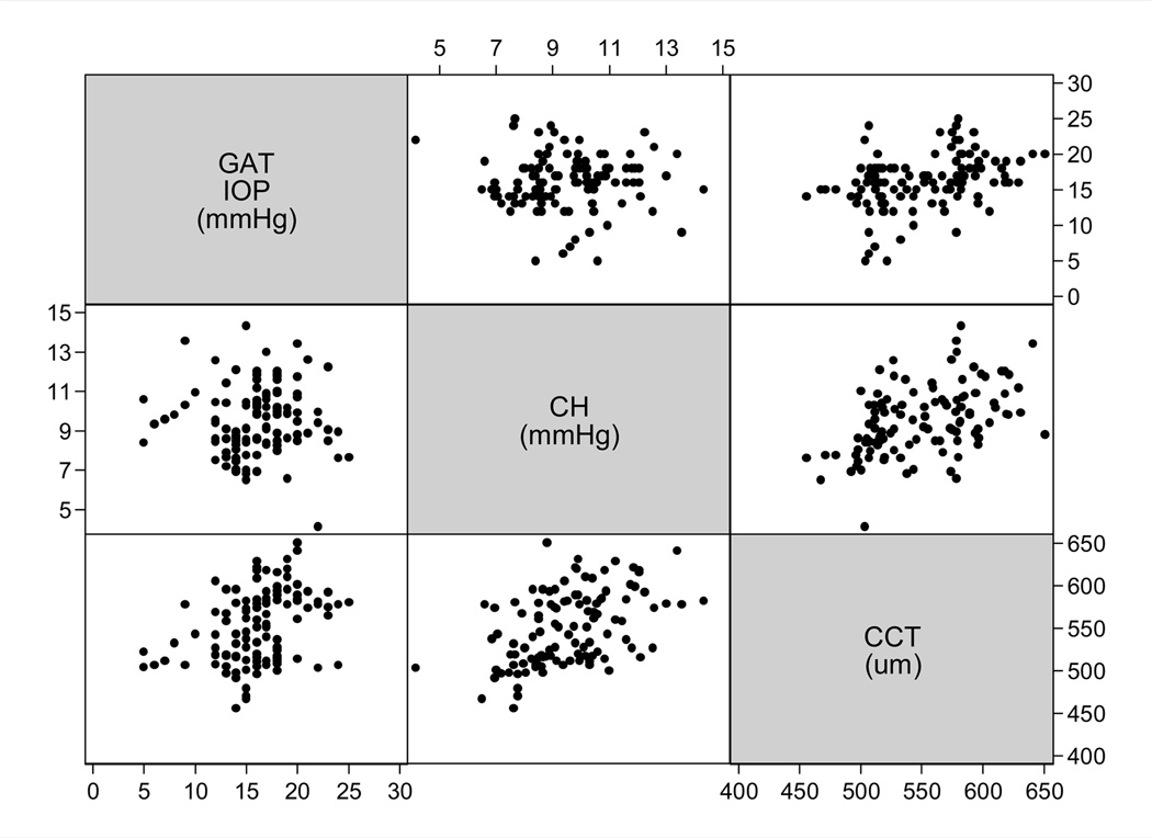
Participants: The study group included 114 eyes of 68 patients with glaucoma followed for an average of 4.0 ± 1.1 years. Visual fields were obtained with standard automated perimetry. Included eyes had a median number of 7 (range, 5-12) tests during follow-up.
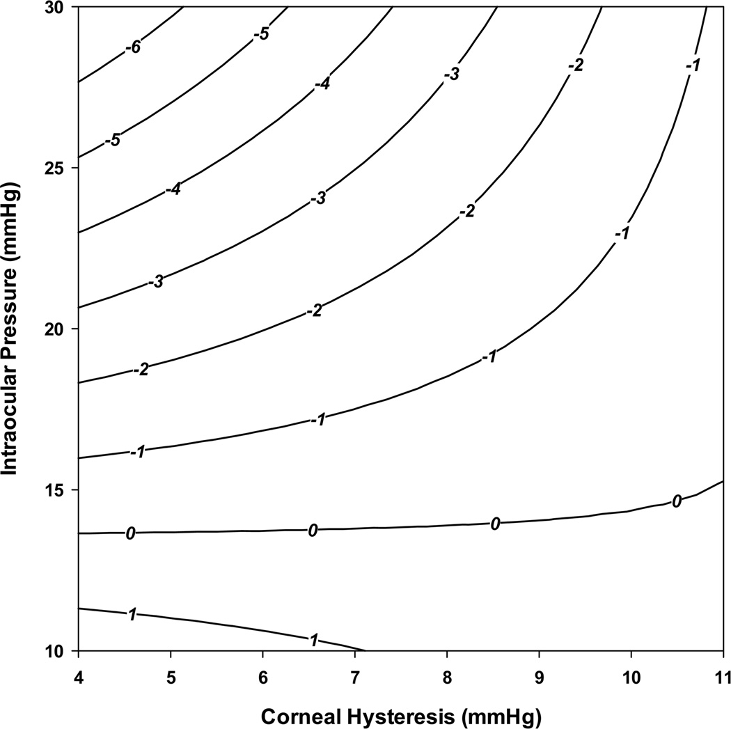
Methods: The CH measurements were acquired at baseline using the Ocular Response Analyzer (Reichert Instruments, Depew, NY). Evaluation of rates of visual field change during follow-up was performed using the visual field index (VFI). Linear mixed models were used to investigate the relationship between rates of visual field loss and baseline CH, baseline intraocular pressure (IOP), and central corneal thickness (CCT), while adjusting for potentially confounding factors. An interaction term between IOP and CH was included in the model to investigate whether the effect of IOP on rates of progression depended on the level of CH.
Main outcome measures: Effects of CH, IOP, and CCT on rates of VFI loss over time.
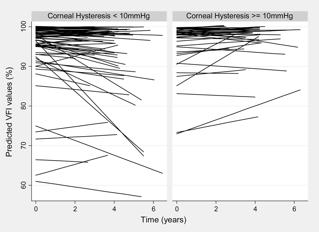
Results: The CH had a significant effect on rates of visual field progression over time. In the univariable model including only CH as a predictive factor along with time and their interaction, each 1 mmHg lower CH was associated with a 0.25%/year faster rate of VFI decline over time (P<0.001). The multivariable model showed that the effect of IOP on rates of progression depended on CH. Eyes with high IOP and low CH were at increased risk for having fast rates of disease progression. The CH explained a larger proportion of the variation in slopes of VFI change than CCT (17.4% vs. 5.2%, respectively).
Conclusions: The CH measurements were significantly associated with risk of glaucoma progression. Eyes with lower CH had faster rates of visual field loss than those with higher CH. The prospective longitudinal design of this study supports the role of CH as an important factor to be considered in the assessment of the risk of progression in patients with glaucoma.
Financial disclosure(s): Proprietary or commercial disclosure may be found after the references.
Copyright © 2013 American Academy of Ophthalmology. Published by Elsevier Inc. All rights reserved.
Figures
Comment in
-
Author reply: To PMID 23642371.Ophthalmology. 2013 Dec;120(12):e85-e86. doi: 10.1016/j.ophtha.2013.09.007. Ophthalmology. 2013. PMID: 24246834 No abstract available.
-
Corneal hysteresis and glaucoma progression.Ophthalmology. 2013 Dec;120(12):e85. doi: 10.1016/j.ophtha.2013.09.008. Ophthalmology. 2013. PMID: 24246835 No abstract available.
References
-
- Collaborative Normal-Tension Glaucoma Study Group. Natural history of normal-tension glaucoma. Ophthalmology. 2001;108:247–253. - PubMed
-
- Heijl A, Bengtsson B, Hyman L, Leske MC. Early Manifest Glaucoma Trial Group. Natural history of open-angle glaucoma. Ophthalmology. 2009;116:2271–2276. - PubMed
-
- Medeiros FA, Zangwill LM, Alencar LM, et al. Rates of progressive retinal nerve fiber layer loss in glaucoma measured by scanning laser polarimetry. Am J Ophthalmol. 2010;149:908–915. - PubMed
-
- Medeiros FA, Susanna R, Jr, Singh K. Who should be treated? In: Weinreb RN, Liebmann J, editors. Medical Treatment of Glaucoma. Amsterdam, The Netherlands: Kugler Publ.; 2010. pp. 1–19.
-
- Leske MC, Heijl A, Hyman L, et al. EMGT Group. Predictors of long-term progression in the Early Manifest Glaucoma Trial. Ophthalmology. 2007;114:1965–1972. - PubMed
Publication types
MeSH terms
Grants and funding
LinkOut - more resources
Full Text Sources
Other Literature Sources
Medical
