

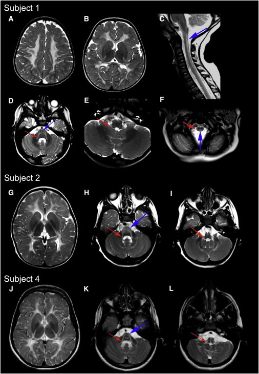
Mutations in DARS cause hypomyelination with brain stem and spinal cord involvement and leg spasticity
- PMID: 23643384
- PMCID: PMC3644624
- DOI: 10.1016/j.ajhg.2013.04.006
Mutations in DARS cause hypomyelination with brain stem and spinal cord involvement and leg spasticity
Abstract
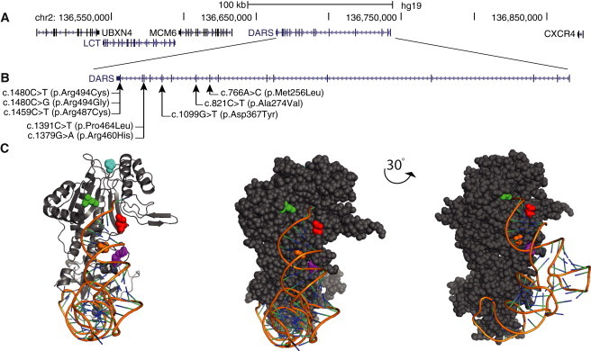
Inherited white-matter disorders are a broad class of diseases for which treatment and classification are both challenging. Indeed, nearly half of the children presenting with a leukoencephalopathy remain without a specific diagnosis. Here, we report on the application of high-throughput genome and exome sequencing to a cohort of ten individuals with a leukoencephalopathy of unknown etiology and clinically characterized by hypomyelination with brain stem and spinal cord involvement and leg spasticity (HBSL), as well as the identification of compound-heterozygous and homozygous mutations in cytoplasmic aspartyl-tRNA synthetase (DARS). These mutations cause nonsynonymous changes to seven highly conserved amino acids, five of which are unchanged between yeast and man, in the DARS C-terminal lobe adjacent to, or within, the active-site pocket. Intriguingly, HBSL bears a striking resemblance to leukoencephalopathy with brain stem and spinal cord involvement and elevated lactate (LBSL), which is caused by mutations in the mitochondria-specific DARS2, suggesting that these two diseases might share a common underlying molecular pathology. These findings add to the growing body of evidence that mutations in tRNA synthetases can cause a broad range of neurologic disorders.
Copyright © 2013 The American Society of Human Genetics. Published by Elsevier Inc. All rights reserved.
Figures
References
-
- van der Knaap M.S., Breiter S.N., Naidu S., Hart A.A., Valk J. Defining and categorizing leukoencephalopathies of unknown origin: MR imaging approach. Radiology. 1999;213:121–133. - PubMed
Publication types
MeSH terms
Substances
Grants and funding
LinkOut - more resources
Full Text Sources
Other Literature Sources
Medical
Molecular Biology Databases
