

Mutations in SCO2 are associated with autosomal-dominant high-grade myopia
- PMID: 23643385
- PMCID: PMC3644634
- DOI: 10.1016/j.ajhg.2013.04.005
Mutations in SCO2 are associated with autosomal-dominant high-grade myopia
Abstract
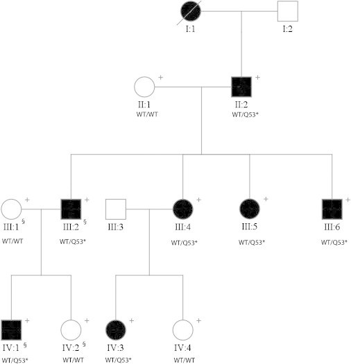
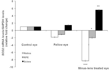
Myopia, or near-sightedness, is an ocular refractive error of unfocused image quality in front of the retinal plane. Individuals with high-grade myopia (dioptric power greater than -6.00) are predisposed to ocular morbidities such as glaucoma, retinal detachment, and myopic maculopathy. Nonsyndromic, high-grade myopia is highly heritable, and to date multiple gene loci have been reported. We performed exome sequencing in 4 individuals from an 11-member family of European descent from the United States. Affected individuals had a mean dioptric spherical equivalent of -22.00 sphere. A premature stop codon mutation c.157C>T (p.Gln53*) cosegregating with disease was discovered within SCO2 that maps to chromosome 22q13.33. Subsequent analyses identified three additional mutations in three highly myopic unrelated individuals (c.341G>A, c.418G>A, and c.776C>T). To determine differential gene expression in a developmental mouse model, we induced myopia by applying a -15.00D lens over one eye. Messenger RNA levels of SCO2 were significantly downregulated in myopic mouse retinae. Immunohistochemistry in mouse eyes confirmed SCO2 protein localization in retina, retinal pigment epithelium, and sclera. SCO2 encodes for a copper homeostasis protein influential in mitochondrial cytochrome c oxidase activity. Copper deficiencies have been linked with photoreceptor loss and myopia with increased scleral wall elasticity. Retinal thinning has been reported with an SC02 variant. Human mutation identification with support from an induced myopic animal provides biological insights of myopic development.
Copyright © 2013 The American Society of Human Genetics. Published by Elsevier Inc. All rights reserved.
Figures

References
-
- Curtin B.J. Harper & Row; Philadelphia: 1985. The Myopias: Basic Science and Clinical Management.
-
- Curtin B.J., Karlin D.B. Axial length measurements and fundus changes of the myopic eye. Am. J. Ophthalmol. 1971;71:42–53. - PubMed
-
- Saw S.M., Gazzard G., Shih-Yen E.C., Chua W.H. Myopia and associated pathological complications. Ophthalmic Physiol. Opt. 2005;25:381–391. - PubMed
-
- Lin L.L., Shih Y.F., Hsiao C.K., Chen C.J., Lee L.A., Hung P.T. Epidemiologic study of the prevalence and severity of myopia among schoolchildren in Taiwan in 2000. J. Formos. Med. Assoc. 2001;100:684–691. - PubMed
-
- Lin L.L., Shih Y.F., Tsai C.B., Chen C.J., Lee L.A., Hung P.T., Hou P.K. Epidemiologic study of ocular refraction among schoolchildren in Taiwan in 1995. Optom. Vis. Sci. 1999;76:275–281. - PubMed
Publication types
MeSH terms
Substances
Grants and funding
LinkOut - more resources
Full Text Sources
Other Literature Sources
Molecular Biology Databases
