. 2013 Dec 10;172(2):579-88.
doi: 10.1016/j.jconrel.2013.04.016. Epub 2013 Apr 30.

Cross-protection by co-immunization with influenza hemagglutinin DNA and inactivated virus vaccine using coated microneedles

Affiliations

Cross-protection by co-immunization with influenza hemagglutinin DNA and inactivated virus vaccine using coated microneedles

Yeu-Chun Kim et al. J Control Release. .

Abstract

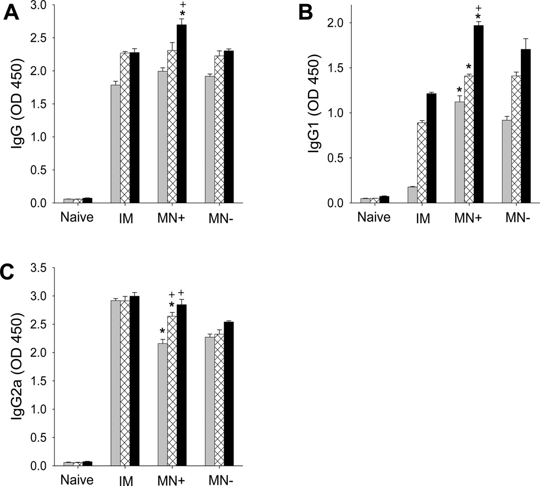
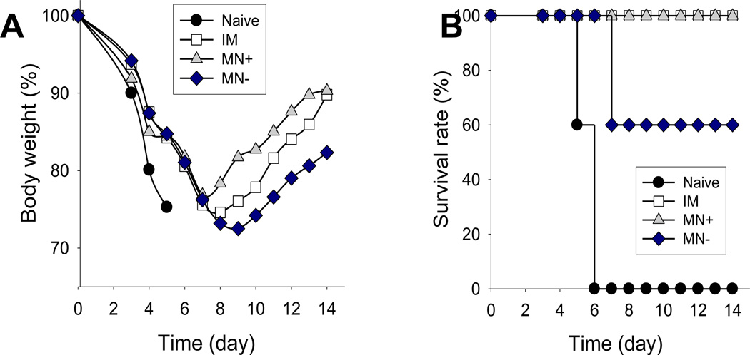
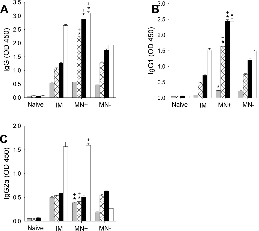
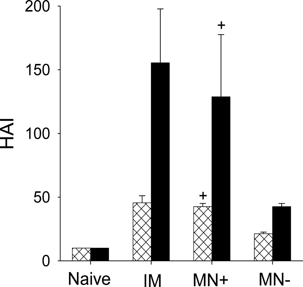
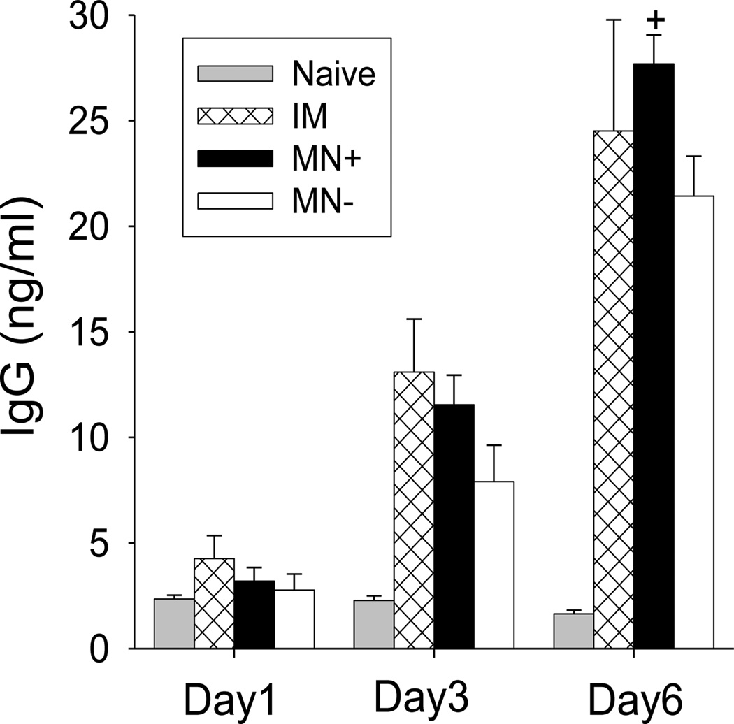
The need for annual revaccination against influenza is a burden on the healthcare system, leads to low vaccination rates and makes timely vaccination difficult against pandemic strains, such as during the 2009 H1N1 influenza pandemic. In an effort toward achieving a broadly protective vaccine that provides cross-protection against multiple strains of influenza, this study developed a microneedle patch to co-immunize with A/PR8 influenza hemagglutinin DNA and A/PR8 inactivated virus vaccine. We hypothesize that this dual component vaccination strategy administered to the skin using microneedles will provide cross-protection against other strains of influenza. To test this hypothesis, we developed a novel coating formulation that did not require additional excipients to increase coating solution viscosity by using the DNA vaccine itself to increase viscosity and thereby enable thick coatings of DNA vaccine and inactivated virus vaccine on metal microneedles. Co-immunization in this way not only generated robust antibody responses against A/PR8 influenza but also generated robust heterologous antibody responses against pandemic 2009 H1N1 influenza in mice. Challenge studies showed complete cross-protection against lethal challenge with live pandemic 2009 H1N1 virus. Control experiments using A/PR8 inactivated influenza virus vaccine with placebo DNA coated onto microneedles produced lower antibody titers and provided incomplete protection against challenge. Overall, this is the first study showing DNA solution as a microneedle coating agent and demonstrating cross-protection by co-immunization with inactivated virus and DNA vaccine using coated microneedles.

Keywords: Coating; Cross-protection; DNA vaccine; Influenza virus; Microneedle.


Conflict of interest statement

The resulting potential conflict of interest has been disclosed and is managed by Georgia Tech and Emory University.

Figures

Figure 1
Figure 1
Microneedles coated with inactivated virus and DNA influenza vaccines. (A) Representative array of five microneedles coated with fluorescein-conjugate BSA shown by bright-field microscopy (scale bar = 800 µm). The coating solution contained 6 mg/ml placebo DNA (B, D, F, H) Fluorescence and (C, E, G, I) bright-field microscopy images of individual microneedles coated with fluorescein-conjugate BSA coated using a coating solution containing (B, C) 2 mg/ml (D, E) 4 mg/ml (F, G) 6 mg/ml (H, I) 8 mg/ml placebo DNA (scale bar = 150 µm).
Figure 2
Figure 2
Effect of trehalose concentration in the coating solution on inactivated virus integrity after coating on microneedles, as assessed by (A) hemagglutination activity and (B) degree of virus aggregation. In the coating solution, inactivated virus concentration was 3mg/ml and DNA concentration was 6 mg/ml in PBS. The dotted line in (B) indicates the size of untreated inactivated virus. Data points represent the average of n = 4 replicates with error bars showing the standard error of the mean (SEM).
Figure 3
Figure 3
Vaccine delivered from microneedles into skin. Representative microscopy images showing individual microneedles coated with fluorescein (green) and Cy3-labeled DNA vaccine (red, but appears yellow here at high concentration) using a formulation containing 3 mg/ml trehalose. Micrographs are shown before (A, C, E) and after (B, D, F) insertion into human cadaver skin for 10 min imaged by (A, B) bright-field and (C, D, E, F) fluorescence microscopy (scale bar = 100µm). Representative histological sections of human cadaver skin imaged by (G) bright-field and (H, I) fluorescence microscopy 10 min after insertion of microneedles coated with (H) Cy3-labeled DNA vaccine and (I) fluorescein (scale bar = 300µm).
Figure 4
Figure 4
PR8 influenza virus-specific antibody responses after vaccination of mice with A/PR8 inactivated virus vaccine and A/PR8 HA-encoding DNA by intramuscular injection (IM), A/PR8 inactivated virus vaccine and A/PR8 HA-encoding DNA by coated microneedles (MN+) and A/PR8 inactivated virus vaccine and placebo DNA by coated microneedles (MN−). The graphs show (A) total serum IgG, (B) IgG1 subtype, (C) IgG2a subtype and (D) IgG2a:IgG1 ratio. Antibody titers were determined 3(■), 5(⊠), 7(■) weeks after immunization. (* comparison with IM, p<0.05, + comparison with MN−, p<0.05, n = 5, ± SEM).
Figure 5
Figure 5
Protection against live-virus challenge after immunization of mice. (A) Body weight change and (B) survival rate after challenge (10×LD50 with pandemic 2009 H1N1 virus). See Fig. 4 caption for details (n = 5).
Figure 6
Figure 6
Pandemic 2009 H1N1 influenza virus-specific antibody responses: (A) total serum IgG, (B) IgG1 subtype, (C) IgG2a subtype and (D) IgG2a:IgG1 ratio. Antibody titers were determined 3(■), 5(⊠), 7(■) weeks after immunization and 4 days (□) after challenge. See Fig. 4 caption for details (* comparison with IM, p<0.05, + comparison with MN−, p<0.05, n = 5, ± SEM).
Figure 7
Figure 7
Hemagglutination inhibition (HAI) titers in sera 7 weeks after immunization (⊠) and 4 days after challenge (■). See Fig. 4 caption for details (+ comparison with MN−, p<0.05, n = 5, ± SEM).
Figure 8
Figure 8
Kinetics of virus-specific IgG antibody production in spleen cells harvested four days after challenge. Spleen cell were cultured on the plates coated with inactivated influenza viral antigen. See Fig. 4 caption for details (+ comparison with MN−, p<0.05, n = 5, ± SEM).

Similar articles

Cited by

References

    1. Neumann G, Noda T, Kawaoka Y. Emergence and pandemic potential of swine-origin H1N1 influenza virus. Nature. 2009;459:931–939. - PMC - PubMed
    1. Gerdil C. The annual production cycle for influenza vaccine. Vaccine. 2003;21:1776–1779. - PubMed
    1. Schwartz B, Gellin B. Vaccination strategies for an influenza pandemic. J. Infect. Dis. 2005;191:1207–1209. - PubMed
    1. Subbarao K, Murphy BR, Fauci AS. Development of effective vaccines against pandemic influenza. Immunity. 2006;24:5–9. - PubMed
    1. Ferguson NM, Cummings DAT, Fraser C, Cajka JC, Cooley PC, Burke DS. Strategies for mitigating an influenza pandemic. Nature. 2006;442:448–452. - PMC - PubMed

Publication types

MeSH terms

Substances