. 2013 May 30;3(5):1580-91.
doi: 10.1016/j.celrep.2013.04.004. Epub 2013 May 2.

Generation of integration-free and region-specific neural progenitors from primate fibroblasts

Affiliations

Generation of integration-free and region-specific neural progenitors from primate fibroblasts

Jianfeng Lu et al. Cell Rep. .

Abstract

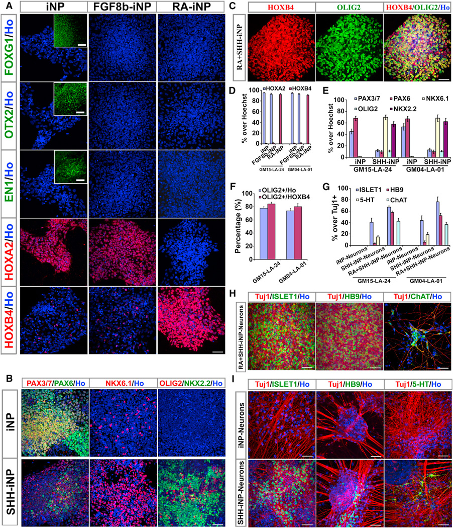
Postnatal and adult human and monkey fibroblasts were infected with Sendai virus containing the Yamanaka factors for 24 hr, then they were cultured in a chemically defined medium containing leukemia inhibitory factor (LIF), transforming growth factor (TGF)-β inhibitor SB431542, and glycogen synthase kinase (GSK)-3β inhibitor CHIR99021 at 39°C for inactivation of the virus. Induced neural progenitor (iNP) colonies appeared as early as day 13 and can be expanded for >20 passages. Under the same defined condition, no induced pluripotent stem cell (iPSC) colonies formed at either 37°C or 39°C. The iNPs predominantly express hindbrain genes and differentiate into hindbrain neurons, and when caudalized, they produced an enriched population of spinal motor neurons. Following transplantation into the forebrain, the iNP-derived cells retained the hindbrain identity. The ability to generate defined, integration-free iNPs from adult primate fibroblasts under a defined condition with predictable fate choices will facilitate disease modeling and therapeutic development.


Figures

Figure 1
Figure 1. Generation of iNPs from Human Fibroblasts
(A) Schematic representation of iNP generation process. (B) Morphological changes during human iNP generation are shown. Fibroblasts were infected with SeV containing OSKM or GFP in the absence (upper row) or presence (middle and lower rows) of LSC. (C) A neural epithelium-like colony was observed in the OSKM+LSC group. (D) Cells from selected colonies were SOX1+ (green), but NANOG− (red). Hoechst staining is blue. (E) Q-PCR of existing SeV genomes in noninfected parent fibroblasts (SeV−, negative control), nine human iNP cell lines, and SeV-infected parent fibroblasts (SeV+, positive control). Data are represented as mean ± SEM. (F) Q-PCR of human ESC markers (OCT4, NANOG, and REX1) during the re-programming process (from the reprogramming experiments of fibroblast cell line GM03815; data from other fibroblast cell lines follow the similar trends, not shown). H9, human ESC line; Days 0–13, days after SeV infection. Data are represented as mean ± SEM. (G) Q-PCR of neural markers (SOX1 and N-Cadherin) during the reprogramming process (from the reprogramming experiments of fibroblast cell line GM03815; data from other fibroblast cell lines follow the similar trends, not shown). Data are represented as mean ± SEM. Scale bars,50 µm (B and D). See also Figure S1 and Tables S1, S2, S5, and S6.
Figure 2
Figure 2. Characterization of Human iNPs
(A) Phase-contrast image for established human iNP cell line 15-LA-24 (passage 5). (B) High-magnification image of cells in the inset in (A). (C–G) Human iNPs were stained positively for NESTIN (C, green), SOX1 (D, red), SOX2 (E, red), FABP7 (F, green), and NOTCH1 (G, green).Hoechst staining is blue. (H) Immunostaining for Ki67 (red). Hoechst staining is blue. (I) RT-PCR shows human iNPs expressed neural progenitor markers: SOX1, SOX2, FABP7, PAX6, and HES5, GAPDH was used as an internal control. RNA was extracted from noninfected parent fibroblasts (Fibroblasts, negative control), three human iNP cell lines (GM15-LA-24, GM15-LA-25, and GM15-LA-01), early passage (passage 5), and late passage (passage 20) of human iNP cell line GM15-LA-24. (J) Growth curve of human iNP cell lines GM15-LA-24, GM15-LA-25, and GM15-LA-01. Cells at passage 5 were used as initial cells. (K) Representative karyotype of human iNP cell line 15-LA-24 at passage 20. Scale bars, 50 mm (A–H). See also Figure S2 and Tables S5 and S6.
Figure 3
Figure 3. In Vitro Differentiation Potential of Human iNPs
(A) Human iNP-derived neural cells showed typical neuronal morphology and expressed markers both for neurons (Tuj1, MAP2, and synapsin) and for astrocytes (GFAP) after 60 days of differentiation. (B) Quantification of MAP2+ neurons and GFAP+ astrocytes among total cells. Data are represented as mean ± SEM. (C) The major population of human iNP-derived neurons was GABA+, and only a small population of neurons was TH+. (D) NKX2.2 (green) and OLIG2 (red) double-positive pre-OPCs were observed in the culture of human iNPs treated with SHH (500 ng/ml) for 1 week. (E) After 10 weeks of differentiation in the glial differentiation medium, O4+ oligodendrocytes were observed in cultures that were treated with SHH (500 ng/ml). (F) Physiological properties of human iNP-derived neurons assessed by whole-cell patch-clamp recordings. (a) Whole-cell recording on a neuron cultured for 10 weeks in vitro. (b) Electrophysiological characteristics of human iNP-derived neurons. (c) Inward Na+ and outward K+ currents were triggered upon −50 to +50 mV voltage steps. The initial currents were enlarged in the inset panel. (d) Action potentials were induced from −60 to +60-pA-injected current steps. (e) Neurons displayed a low rate of spontaneous action potential spiking with a subthreshold oscillatory potential (−30 mV). (f) Both excitatory (inward current) and inhibitory (outward current) spontaneous postsynaptic currents were recorded when neurons were held at 0 mV. The excitatory responses were eliminated after treatment with CNQX, and the inhibitory responses were further eliminated upon the presence of additive bicuculline in the extracellular solution. The asterisks indicate individual excitatory (sEPSCs) and inhibitory (sIPSCs) events. Scale bars, 50 mm (A, C, D, and E). See also Figure S3 and Table S6.
Figure 4
Figure 4. Generation and Characterization of iNPs from Monkey Fibroblasts
(A) A SOX1+ (green) and NANOG− (red) neuroepithelial colony was observed in the reprogramming culture from monkey fibroblasts. (B) Phase-contrast image of monkey iNPs. Inset is the high-magnification image. (C–F) Monkey iNPs were NESTIN+ (C, green), SOX1+ (D, red), SOX2+ (E, red), and FABP7+ (F, green). (G) Immunostaining for Ki67 (red). (H) Growth curve from monkey iNP cell lines (47-LA-01, 47-LA-05, 47-LA-11)and from human iNP cell lines (GM15-LA-24, GM15-LA-24, GM15-LA-24). Cells at passage 5 were used as initial cells. (I) Q-PCR of existing SeV genomes in noninfected parent fibroblasts (Fibroblast, negative control), three monkey iNP cell lines (47-LA-01, 47-LA-05, 47-LA-11), and SeV-infected human fibroblasts (SeV, positive control). Data are represented as mean ± SEM. (J) Monkey iNP-derived neural cells showed typical neuronal morphology and expressed markers both for neurons (Tuj1, MAP2, and synapsin) and for astrocytes (GFAP) after 8 weeks of differentiation. O4+ oligodendrocytes were observed in differentiation cultures from ventralized monkey iNPs. (K) Physiological properties of monkey iNP-derived neurons assessed by whole-cell patch-clamp recordings. (a) Whole-cell recording on a neuron at 10 weeks in vitro. (b) Electrophysiological characteristics of monkey iNP-derived neurons. (c) Inward Na+ and outward K+ currents were triggered upon −50 to +50 mV voltage steps. The initial currents were enlarged in the inset panel. (d) Action potentials were induced from −60 to +60-pA-injected current steps. (e) Neurons displayed a low rate of spontaneous action potential spiking with a subthreshold oscillatory potential (−30 mV). (f) Both excitatory (inward current) and inhibitory (outward current) spontaneous postsynaptic currents were recorded when neurons were held at 0 mV. The excitatory responses were eliminated after treatment with CNQX, and the inhibitory responses were further eliminated upon the presence of additive bicuculline in extracellular solution. The asterisks indicate individual excitatory (sEPSCs) and inhibitory (sIPSCs) events. Scale bars, 50 µm (A–G and J).
Figure 5
Figure 5. Identity and Generation of Region-Specific Progenitors and Neurons from iNPs
(A) Human iNPs predominantly expressed hindbrain marker HOXA2, and only a small portion of cells expressed marker for posterior hindbrain or spinal cord (HOXB4), but not anterior markers FOXG1, OTX2, and EN1. When treated with FGF8b (100 ng/ml, FGF8b-iNP), the regional identity was not altered. When treated with RA (0.1 µM, RA-iNP), human iNPs were posteriorly patterned and predominantly expressed marker for posterior hindbrain or cervical spinal cord (HOXB4). Insets show positive controls for FOXG1, OTX2, and EN1, respectively. (B) Human iNPs predominantly expressed marker for dorsal (PAX3/PAX7) and marker for middle part of hindbrain (PAX6), and only a small portion of cells expressed marker for ventral hindbrain (NKX6.1). When treated with SHH (1,000 ng/ml, SHH-iNP), human iNPs were ventrally patterned and predominantly expressed markers for ventral hindbrain (NKX6.1 and NKX2.2). (C) When treated with RA (0.1 µM) and SHH (500 ng/ml) (RA+SHH-iNP), most human iNP cells became OLIG2+ and HOXB4+ spinal cord motor neuron precursors. (D) Quantification of cells expressing HOXA2 and HOXB4 in (A). Data are represented as mean ± SEM. (E) Quantification of cells in (B). Data are represented as mean ± SEM. (F) Quantification of cells in (C). Data are represented as mean ± SEM. (G) Quantification of cells in (H) and (I). Data are represented as mean ± SEM. (H) The major neuronal population in the RA and SHH-treated iNP cultures (RA+SHH-iNP-Neurons) was HB9+, ISLET1+, and ChAT+ spinal cord motor neurons. (I) The ventralized neural progenitors differentiated to ISLET1+, HB9+, and 5-HT+ ventral neurons (SHH-iNP-Neurons). Cell nuclei were stained with Hoechst (blue). Scale bars, 50 µm (A–C, H, and I). See also Figure S4 and Tables S3 and S6.
Figure 6
Figure 6. In Vivo Differentiation Potential of Human iNPs
(A, C, E, G, and I) One month posttransplantation, human cells, stained for STEM121, were present in the lateral ventricle (A) and were positive for Tuj1 (C), GFAP (E) but rarely for NeuN (G), and not for MBP (I). (B, D, F, H, and J) Four months posttransplantation, the human cells were also in parenchyma tissues near the ventricle (B) and were positive for Tuj1 (D), GFAP (F), NeuN (H, arrowheads), and MBP (J, arrows indicate MBP+ fibers). (K–N) Four months posttransplantation, the human cells were detected as FOXG1- (K), OTX2-(L), HOXA2+ (M), and HOXB4+ (N). Nuclei were stained with Hoechst (blue). Scale bars, 200 µm (A and B) and 50 µm (C–N). See also Figure S5 and Table S6.

References

    1. Ban H, Nishishita N, Fusaki N, Tabata T, Saeki K, Shikamura M, Takada N, Inoue M, Hasegawa M, Kawamata S, Nishikawa S. Efficient generation of transgene-free human induced pluripotent stem cells (iPSCs) by temperature-sensitive Sendai virus vectors. Proc. Natl. Acad. Sci. USA. 2011;108:14234–14239. - PMC - PubMed
    1. Brambrink T, Foreman R, Welstead GG, Lengner CJ, Wernig M, Suh H, Jaenisch R. Sequential expression of pluripotency markers during direct reprogramming of mouse somatic cells. Cell Stem Cell. 2008;2:151–159. - PMC - PubMed
    1. Caiazzo M, Dell’Anno MT, Dvoretskova E, Lazarevic D, Taverna S, Leo D, Sotnikova TD, Menegon A, Roncaglia P, Colciago G, et al. Direct generation of functional dopaminergic neurons from mouse and human fibroblasts. Nature. 2011;476:224–227. - PubMed
    1. Efe JA, Hilcove S, Kim J, Zhou H, Ouyang K, Wang G, Chen J, Ding S. Conversion of mouse fibroblasts into cardiomyocytes using a direct reprogramming strategy. Nat. Cell Biol. 2011;13:215–222. - PubMed
    1. Ferrari S, Griesenbach U, Iida A, Farley R, Wright AM, Zhu J, Munkonge FM, Smith SN, You J, Ban H, et al. Sendai virus-mediated CFTR gene transfer to the airway epithelium. Gene Ther. 2007;14:1371–1379. - PubMed

Publication types

MeSH terms