. 2013 May 30;3(5):1389-97.
doi: 10.1016/j.celrep.2013.03.040. Epub 2013 May 2.

Chromosomal instability triggered by Rrm2b loss leads to IL-6 secretion and plasmacytic neoplasms

Affiliations

Chromosomal instability triggered by Rrm2b loss leads to IL-6 secretion and plasmacytic neoplasms

Lufen Chang et al. Cell Rep. .

Abstract

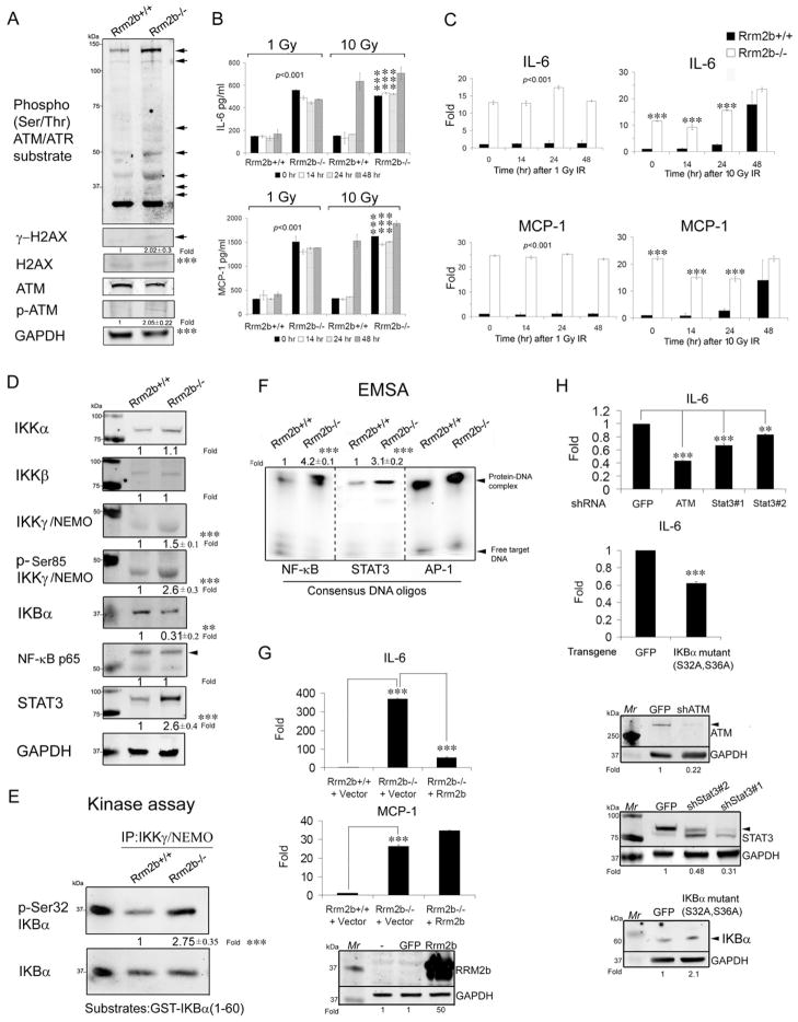
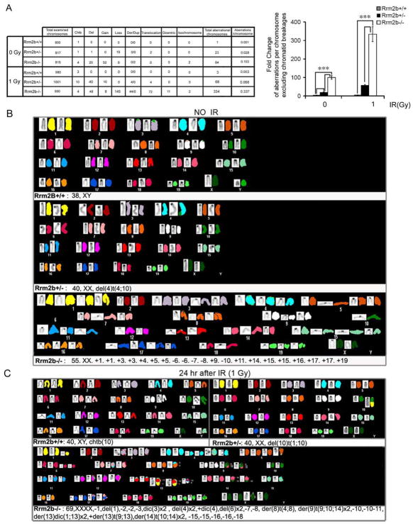
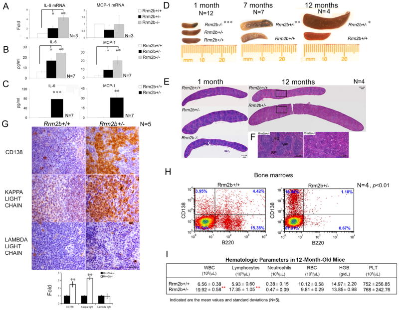
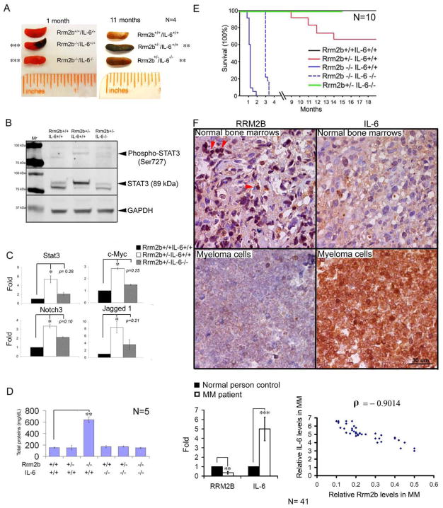
Chronic inflammation has a tight cause-and-effect relationship with DNA damage by inflicting tissue damage and increasing cancer risk. Rrm2b, a key enzyme in de novo deoxyribonucleotide synthesis, is involved in DNA damage repair, but its role in cancer development has yet to be demonstrated. In this work, Rrm2b gene loss led to severe numerical and structural chromosome abnormalities that caused ATM activation, inducing p-Ser85 IKKγ/NEMO and IκB kinase (IKK). NF-κB consequently induced by IKK triggered sustained IL-6 expression that constitutively activated STAT3 in Rrm2b-deficient cells. High plasma interleukin-6 (IL-6) and associated hematologic disorders were observed in Rrm2b-/- mice, and 30%-40% of aged Rrm2b heterozygous knockout mice developed plasma cell neoplasms and suffered from progressive splenomegaly and ascites. The genetic ablation of IL-6 suppressed STAT3 induction and delayed disease onset in Rrm2b-/- mice, extending their lifespan. Thus, Rrm2b plays a crucial role in maintaining chromosomal stability and preventing chronic-inflammation-associated tumorigenesis.


Figures

Figure 1
Figure 1. Sustained IL-6 expression mediated by constitutive NF-κB and STAT3 in Rrm2b-deficient Cells
(A) Immunoblotting (IB) analysis of ATM-related signaling proteins in MEFs derived from Rrm2b+/+ and Rrm2b−/− E14.5 embryos. Phosphorylated H2Ax at Ser139, γ-H2AX; Phosphorylated ATM at Ser1981, p-ATM. (B) IL-6 and MCP-1 cytokines from Rrm2b+/+ and Rrm2b−/− MEF media before and after 1 or 10 gray (Gy) IR at the indicated time-points were analyzed by Luminex multiplex cytokine assays. (C) RT-qPCR analysis of IL-6 and MCP-1 mRNAs of MEFs before and after 1 or 10 Gy IR. (D) IB analysis of NF-KB and STAT3 signaling proteins in MEFs from each genotype. (E) IKK kinase activity in MEFs of each genotype analyzed by the IKKγ–immunoprecipitated complex on GST-IKBα(1-60) recombinant proteins followed by IB with p-Ser32 IKBα and IKBα antibodies. (F) NF-kB and STAT3 DNA binding activity in MEFs from each genotype analyzed by a non-radioactive electrophoretic mobility shift assay (EMSA) after quantification and normalization with AP-1 binding activity, which served as a control for extract quality and loading. (G) RT-qPCR analysis of IL-6 and MCP-1 mRNAs in Rrm2b−/− fibroblasts after infection with lentiviral Rrm2b transgenes. Transgene proteins were examined by IB in the bottom panel. (H) RT-qPCR analysis of IL-6 mRNA in Rrm2b−/− MEFs after infection with lentiviruses expressing shRNAs against the indicated target genes or GFP (control). Knocked-down proteins were analyzed by IB and shown in the bottom panels. The fold-chang (Fold) in IB, kinase activity, and EMSA assays is a relative value to WT control after subtraction from the loading control, which given was given an arbitrary value of 1.0. Data represented as mean ± standard deviation (SD) were average from three independent MEFs derived from different embryos for each genotype. **,p< 0.01; ***,p< 0.001. See also Figure S1.
Figure 2
Figure 2. Increased Numerical and Structural Chromosome Abnormalities in Rrm2b Deficient Cells
(A) The values in the Table represent the number of each different type of aberrant chromosomes in the examined total chromosomes from three independent cell lines before and 24 h after 1Gy IR for each genotype analyzed by FISH karyotype analysis. The fold-chang of chromosome aberration frequencies excluding Chtb among the genotypes was shown in the histograms. Chromatid break, Chtb; Deletion, del; Extra copies of chromosomes, gain; Missing copies of chromosomes, loss; Derivative chromosome, der; Duplicated chromosome, dup; Chromosomes having two centromeres, dicentric; Fusion of chromosomes, isochromosome. A representative pseudocolor spectral karyotype image of one MEF from each genotype before (B) and 24 h after 1 Gy IR (C). Gain, +; Loss, −; Translocation, t; Dicentric, dic. Data represented as mean ± SD. ***,p< 0.001. See also Figure S2.
Figure 3
Figure 3. Elevated IL-6 and MCP-1 and the Plasma Cell Neoplasia Phenotype in Aged Rrm2b+/− mice
IL-6 and MCP-1 (A) mRNAs in 1-month-old bone marrows from each genotype analyzed by qPCR. IL-6 and MCP-1 protein levels from blood serum of 1-month-old (B) and from 9- to 12-month-old mice (C). (D) Spleens from each genotype at the indicated ages. The data shown were obtained from a litter-matched group of mice. (E) Tiling images of representative H&E stained sections of spleen from each genotype at 1 and 12 months. Higher magnification of the boxed area of 12-month-old spleens is shown in (F) Marginal Zone, MG; White pulp, WP; Red pulp, RP. (G) A representative section of IHC staining for CD138, KAPPA light chain and LAMBDA light chain antibodies (Brown color) from 12-month-old Rrm2b+/+ and Rrm2b+/− spleens. (H) Fluorescence-activated cell sorting (FACS) of cells derived from Rrm2b+/+ and Rrm2b+/− primary bone marrows for plasma cells using CD138 and B220 antibodies. (I) CBC of peripheral blood from 12-month-old mice. White blood cell, WBC; Red blood cell, RBC; Hemoglobin, HBG; Platelet, PLT. Data represented as mean ± SD. *,p< 0.05; **,p< 0.01; ***,p< 0.001. See also Figure S3.
Figure 4
Figure 4. IL-6 Deficiency Delayed Disease Onset and Mortality of Rrm2b−/− mice
(A) Absence of splenic hemorrhage in the representative 1-month-old Rrm2b−/−IL-6−/− spleen and 11-month-old Rrm2b+/−IL-6−/− spleen compared to their age-matched littermates. (B) Analysis of STAT3 activity and protein expression in 12-month-oldbone marrows with indicated genotypes by IB analysis using antibodies against phospho-STAT3 (Ser727) and STAT3. (C) mRNA levels of the indicated genes in 12-month-old bone marrows with indicated genotypes. (D) Deletion of IL-6 blocked urinary protein secretion in Rrm2b−/− mice at 5 weeks of age. (E) Kaplan-Meier survival curve of mice with indicated genotypes. (F) A representative section of IHC staining for Rrm2b and IL-6 antibodies (Brown color) from normal human bone marrows and myeloma cells of a MM patient (N=41). Nuclear Rrm2b indicated by arrow heads. The quantification of Rrm2b and IL-6 signals was shown in the histogram. Correlation between relative IL-6 and Rrm2b levels in MM patients was plotted and analyzed by Pearson correlation coefficient analyses. ρ denotes Pearson’s correlation. Data represented as mean ± SD. *,p< 0.05; **,p< 0.01; ***,p< 0.001. See also Figure S4.

Similar articles

Cited by

References

    1. Bourdon A, Minai L, Serre V, Jais JP, Sarzi E, Aubert S, Chretien D, de Lonlay P, Paquis-Flucklinger V, Arakawa H, et al. Mutation of RRM2B, encoding p53-controlled ribonucleotide reductase (p53R2), causes severe mitochondrial DNA depletion. Nature genetics. 2007;39:776–780. - PubMed
    1. Chang L, Zhou B, Hu S, Guo R, Liu X, Jones SN, Yen Y. ATM-mediated serine 72 phosphorylation stabilizes ribonucleotide reductase small subunit p53R2 protein against MDM2 to DNA damage. Proceedings of the National Academy of Sciences of the United States of America. 2008;105:18519–18524. - PMC - PubMed
    1. Grivennikov S, Karin M. Autocrine IL-6 signaling: a key event in tumorigenesis? Cancer Cell. 2008;13:7–9. - PubMed
    1. Grivennikov SI, Greten FR, Karin M. Immunity, inflammation, and cancer. Cell. 2010;140:883–899. - PMC - PubMed
    1. Kimura T, Takeda S, Sagiya Y, Gotoh M, Nakamura Y, Arakawa H. Impaired function of p53R2 in Rrm2b-null mice causes severe renal failure through attenuation of dNTP pools. Nature genetics. 2003;34:440–445. - PubMed

Publication types

MeSH terms

LinkOut - more resources