

Sleep-disordered breathing: effects on brain structure and function
- PMID: 23643610
- PMCID: PMC3778068
- DOI: 10.1016/j.resp.2013.04.021
Sleep-disordered breathing: effects on brain structure and function
Abstract
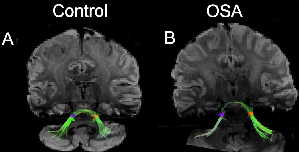
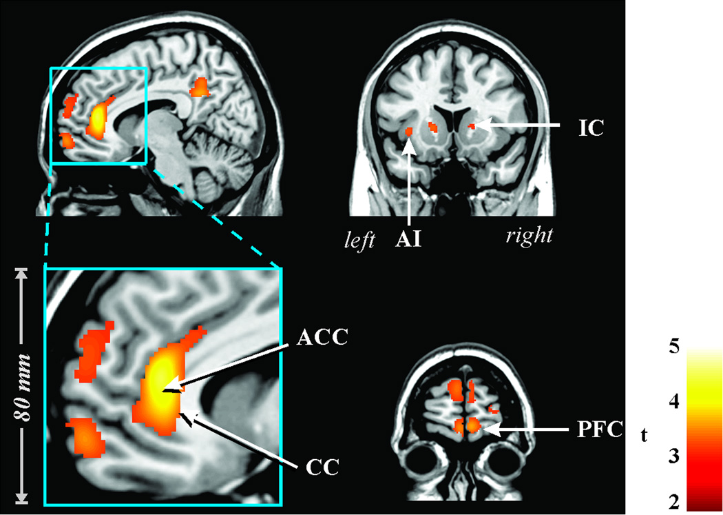
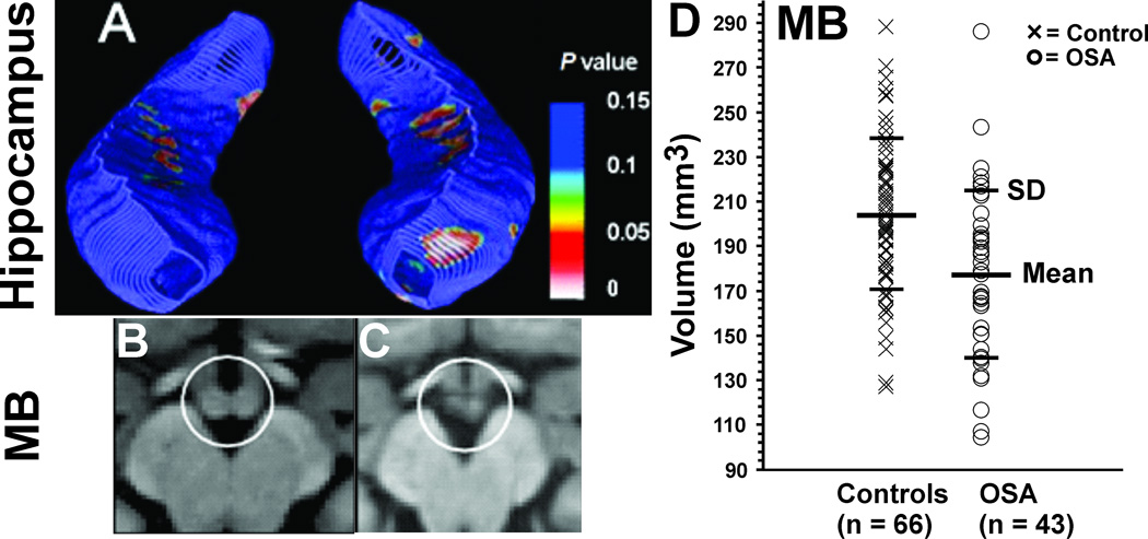
Sleep-disordered breathing is accompanied by neural injury that affects a wide range of physiological systems which include processes for sensing chemoreception and airflow, driving respiratory musculature, timing circuitry for coordination of breathing patterning, and integration of blood pressure mechanisms with respiration. The damage also occurs in regions mediating emotion and mood, as well as areas regulating memory and cognitive functioning, and appears in structures that serve significant glycemic control processes. The injured structures include brain areas involved in hormone release and action of major neurotransmitters, including those playing a role in depression. The injury is reflected in a range of structural magnetic resonance procedures, and also appears as functional distortions of evoked activity in brain areas mediating vital autonomic and breathing functions. The damage is preferentially unilateral, and includes axonal projections; the asymmetry of the injury poses unique concerns for sympathetic discharge and potential consequences for arrhythmia. Sleep-disordered breathing should be viewed as a condition that includes central nervous system injury and impaired function; the processes underlying injury remain unclear.
Keywords: Autonomic; Congenital central hypoventilation; Dyspnea; Hypoxia; Magnetic resonance imaging; Neural injury; Obstructive sleep apnea.
Copyright © 2013 Elsevier B.V. All rights reserved.
Figures
References
-
- Allen AM, Moeller I, Jenkins TA, Zhuo J, Aldred GP, Chai SY, Mendelsohn FA. Angiotensin receptors in the nervous system. Brain research bulletin. 1998;47:17–28. - PubMed
-
- Banzett RB, Mulnier HE, Murphy K, Rosen SD, Wise RJ, Adams L. Breathlessness in humans activates insular cortex. Neuroreport. 2000;11:2117–2120. - PubMed
-
- Berry RB, White DP, Roper J, Pillar G, Fogel RB, Stanchina M, Malhotra A. Awake negative pressure reflex response of the genioglossus in OSA patients and normal subjects. J Appl Physiol. 2003;94:1875–1882. - PubMed
-
- Buckley MJ, Charles DP, Browning PG, Gaffan D. Learning and retrieval of concurrently presented spatial discrimination tasks: role of the fornix. Behav Neurosci. 2004;118:138–149. - PubMed
-
- Chen ML, Witmans MB, Tablizo MA, Jubran RF, Turkel SB, Tavare CJ, Keens TG. Disordered respiratory control in children with partial cerebellar resections. Pediatr Pulmonol. 2005;40:88–91. - PubMed
Publication types
MeSH terms
Substances
Grants and funding
LinkOut - more resources
Full Text Sources
Other Literature Sources
Medical
