

Altered nucleolar morphology in substantia nigra dopamine neurons following 6-hydroxydopamine lesion in rats
- PMID: 23643997
- PMCID: PMC3679339
- DOI: 10.1016/j.neulet.2013.04.033
Altered nucleolar morphology in substantia nigra dopamine neurons following 6-hydroxydopamine lesion in rats
Abstract
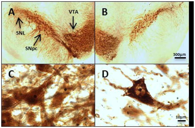
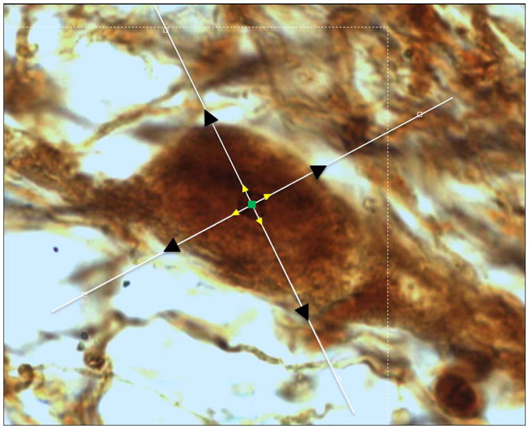
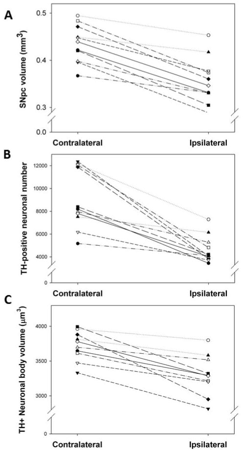
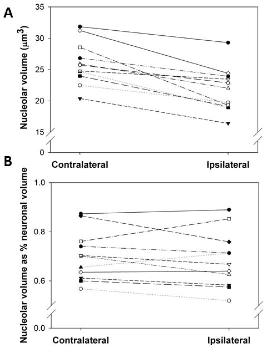
The nucleolus, the site of ribosomal ribonucleic acid (rRNA) transcription and assembly, is an important player in the cellular response to stress. Altered nucleolar function and morphology, including decreased nucleolar volume, has been observed in Parkinson's disease; thus the nucleolus represents a potential indicator of neurodegeneration in the disease. This study determined the effects of a partial unilateral intrastriatal 6-hydroxydopamine (6-OHDA) lesion, which models the dopaminergic loss found in Parkinson's disease, on the nucleoli of dopaminergic cells in the substantia nigra pars compacta (SNpc). Adult male Long-Evans rats underwent unilateral intrastriatal infusion of 6-OHDA (12.5μg). Lesions were verified by amphetamine-stimulated rotation 7 days later, and rats were euthanized 14 days after infusion. Coronal sections (50μm) were stained for tyrosine hydroxylase-silver nucleolar (TH-AgNOR) stain using MultiBrain Technology (NeuroScience Associates), which resulted in clearly defined nucleoli and neuronal outlines. Stereological methods were used to compare dopaminergic morphology between lesioned and intact hemispheres in each rat. In cells exhibiting a definable nucleolus, nucleolar volume was decreased by 16% on the ipsilateral side. The ipsilateral SNpc also exhibited an 18% decrease in SNpc planimetric volume, a 46% decrease in total TH-positive neuron number, and an 11% decrease in neuronal body volume (all P<0.05 by paired t-test). These findings suggest that the 6-OHDA lesion alters nucleolar morphology and that these changes are similar to those occurring in Parkinson's disease.
Copyright © 2013 Elsevier Ireland Ltd. All rights reserved.
Figures
Similar articles
-
Differential effects of intrastriatal 6-hydroxydopamine on cell number and morphology in midbrain dopaminergic subregions of the rat.Brain Res. 2014 Jul 29;1574:113-9. doi: 10.1016/j.brainres.2014.05.045. Epub 2014 Jun 9. Brain Res. 2014. PMID: 24924804 Free PMC article.
-
A novel use of combined tyrosine hydroxylase and silver nucleolar staining to determine the effects of a unilateral intrastriatal 6-hydroxydopamine lesion in the substantia nigra: a stereological study.J Neurosci Methods. 2012 Sep 30;210(2):187-94. doi: 10.1016/j.jneumeth.2012.07.013. Epub 2012 Jul 28. J Neurosci Methods. 2012. PMID: 22850559 Free PMC article.
-
Fibroblast growth factor-20 protects against dopamine neuron loss in vitro and provides functional protection in the 6-hydroxydopamine-lesioned rat model of Parkinson's disease.Neuropharmacology. 2012 Dec;63(7):1268-77. doi: 10.1016/j.neuropharm.2012.07.029. Epub 2012 Aug 10. Neuropharmacology. 2012. PMID: 22971544
-
Relationship between microglial activation and dopaminergic neuronal loss in the substantia nigra: a time course study in a 6-hydroxydopamine model of Parkinson's disease.J Neurochem. 2009 Aug;110(3):966-75. doi: 10.1111/j.1471-4159.2009.06189.x. Epub 2009 Jun 22. J Neurochem. 2009. PMID: 19549006
-
Neonatal 6-hydroxydopamine lesioning of rats and dopaminergic neurotoxicity: proposed animal model of Parkinson's disease.J Neural Transm (Vienna). 2022 Jun;129(5-6):445-461. doi: 10.1007/s00702-022-02479-4. Epub 2022 Mar 12. J Neural Transm (Vienna). 2022. PMID: 35279767 Review.
Cited by
-
Impaired ribosome biogenesis: mechanisms and relevance to cancer and aging.Aging (Albany NY). 2019 Apr 26;11(8):2512-2540. doi: 10.18632/aging.101922. Aging (Albany NY). 2019. PMID: 31026227 Free PMC article. Review.
-
Genetic mutations linked to Parkinson's disease differentially control nucleolar activity in pre-symptomatic mouse models.Dis Model Mech. 2017 May 1;10(5):633-643. doi: 10.1242/dmm.028092. Epub 2017 Mar 30. Dis Model Mech. 2017. PMID: 28360124 Free PMC article.
-
Coronin 2B deficiency induces nucleolar stress and neuronal apoptosis.Cell Death Dis. 2024 Jun 27;15(6):457. doi: 10.1038/s41419-024-06852-x. Cell Death Dis. 2024. PMID: 38937439 Free PMC article.
-
Alcohol exposure suppresses ribosome biogenesis and causes nucleolar stress in cranial neural crest cells.PLoS One. 2024 Jun 28;19(6):e0304557. doi: 10.1371/journal.pone.0304557. eCollection 2024. PLoS One. 2024. PMID: 38941348 Free PMC article.
-
The nucleolar protein nucleophosmin is essential for autophagy induced by inhibiting Pol I transcription.Sci Rep. 2015 Mar 10;5:8903. doi: 10.1038/srep08903. Sci Rep. 2015. PMID: 25754892 Free PMC article.
References
-
- Bethel-Brown CS, Zhang H, Fowler SC, Chertoff ME, Watson GS, Stanford JA. Within-session analysis of amphetamine-elicited rotation behavior reveals differences between young adult and middle-aged F344/BN rats with partial unilateral striatal dopamine depletion. Pharmacol Biochem Behav. 2010;96:423–428. - PMC - PubMed
-
- Carvalho GA, Nikkhah G. Subthalamic nucleus lesions are neuroprotective against terminal 6-OHDA-induced striatal lesions and restore postural balancing reactions. Exp Neurol. 2001;171:405–417. - PubMed
-
- Cheng HW, Tong J, McNeill TH. Lesion-induced axon sprouting in the deafferented striatum of adult rat. Neurosci Lett. 1998;242:69–72. - PubMed
Publication types
MeSH terms
Substances
Grants and funding
LinkOut - more resources
Full Text Sources
Other Literature Sources
Miscellaneous
