. 2013 May 23;497(7450):507-11.
doi: 10.1038/nature12105. Epub 2013 May 5.

Lamin A/C and emerin regulate MKL1-SRF activity by modulating actin dynamics

Affiliations

Lamin A/C and emerin regulate MKL1-SRF activity by modulating actin dynamics

Chin Yee Ho et al. Nature. .

Abstract

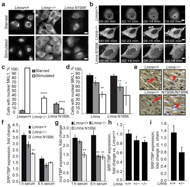
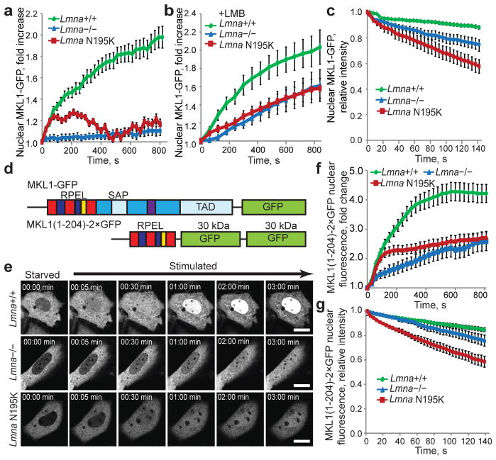
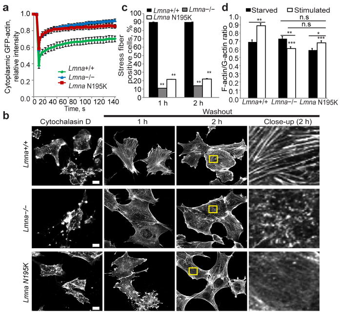
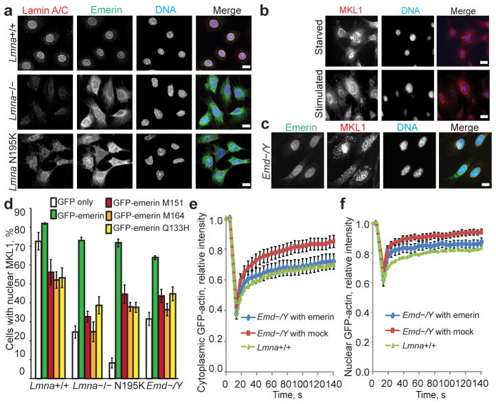
Laminopathies, caused by mutations in the LMNA gene encoding the nuclear envelope proteins lamins A and C, represent a diverse group of diseases that include Emery-Dreifuss muscular dystrophy (EDMD), dilated cardiomyopathy (DCM), limb-girdle muscular dystrophy, and Hutchison-Gilford progeria syndrome. Most LMNA mutations affect skeletal and cardiac muscle by mechanisms that remain incompletely understood. Loss of structural function and altered interaction of mutant lamins with (tissue-specific) transcription factors have been proposed to explain the tissue-specific phenotypes. Here we report in mice that lamin-A/C-deficient (Lmna(-/-)) and Lmna(N195K/N195K) mutant cells have impaired nuclear translocation and downstream signalling of the mechanosensitive transcription factor megakaryoblastic leukaemia 1 (MKL1), a myocardin family member that is pivotal in cardiac development and function. Altered nucleo-cytoplasmic shuttling of MKL1 was caused by altered actin dynamics in Lmna(-/-) and Lmna(N195K/N195K) mutant cells. Ectopic expression of the nuclear envelope protein emerin, which is mislocalized in Lmna mutant cells and also linked to EDMD and DCM, restored MKL1 nuclear translocation and rescued actin dynamics in mutant cells. These findings present a novel mechanism that could provide insight into the disease aetiology for the cardiac phenotype in many laminopathies, whereby lamin A/C and emerin regulate gene expression through modulation of nuclear and cytoskeletal actin polymerization.


Conflict of interest statement

Competing financial interests

The authors declare no competing financial interests.

Figures

Figure 1
Figure 1. Impaired nuclear translocation of MKL1 in lamin A/C-deficient and Lmna N195K mutant cells
(a) Lmna−/− and Lmna N195K MEFs had a lower fraction of nuclear MKL1 after serum stimulation than Lmna+/+ cells, based on MKL1 immunofluorescence. Scale bar, 10 μm. (b) Time-lapse sequences of cells expressing MKL1-GFP stimulated with serum (see Suppl. Information for videos). Scale bar, 10 μm. (c) Quantitative analysis of MEFs with positive nuclear MKL1 staining in response to serum stimulation (N 50 per cell line). (d) Quantitative analysis of myocytes with nuclear MKL1 in cardiac sections from Lmna−/− and LmnaN195K/N195K mice as well as littermate controls (N = 3 for each). (e) Representative histological cardiac tissue sections from Lmna−/− and LmnaN195K/N195K mice and age-matched wild-type littermates stained for MKL1 (brown). Red arrows denote example of MKL1-positive nucleus; arrow head denotes an MKL1-negative nucleus. Scale bar, 20 μm. (fg) Gene expression of serum response factor (SRF) and vinculin (Vcl) in Lmna+/+, Lmna −/− and Lmna N195K MEFs after 1 h and 6 h of serum stimulation. Values were based on 3 independent experiments and were normalized to TATA binding protein (TBP). (h) Gene expression of SRF in Lmna+/+ (N = 9), Lmna+/ (N = 11) and Lmna −/− (N = 10) cardiac tissue. Values were normalized to TBP. (i) Gene expression of SRF in Lmna+/+ (N = 5) and Lmna+/ (N = 7) cardiac tissue collected 1 week after transverse aortic constriction (TAC) surgery. Values were normalized to TBP and compared to those from sham animals. Statistical significance determined by Student’s t-test, compared to Lmna+/+ MEFs; *, indicates P ≤ 0.05; **, indicates P ≤ 0.01; ****, indicates P ≤ 0.0001. Error bars, s.e.m.
Figure 2
Figure 2. Changes in nuclear import and export are specific to MKL1 and are caused by altered actin dynamics in Lmna−/− and Lmna N195K cells
(a, b) Change in nuclear fluorescence intensity over time upon serum stimulation in Lmna+/+, Lmna−/− and Lmna N195K MEFs expressing MKL1-GFP in the absence (a) or presence of leptomycin B (b). Values were normalized to the initial nuclear fluorescence intensity before serum addition. N = 20 for each cell line. (c) Fluorescence loss in photobleaching (FLIP) experiments of MKL1-GFP to measure nuclear export. Increased loss of nuclear fluorescence indicates a higher rate of nuclear export of MKL1-GFP in lamin mutant cells. N = 10 for each cell line. (d) Schematic representation (not drawn to scale) of full length MKL1-GFP (top) and MKL1(1–204)–2×GFP (bottom), consisting of the N-terminal actin binding domain of MKL1 fused to two GFP moieties. RPEL motifs depicted in blue and red, NLS in yellow, DNA binding domain (SAP) and transcriptional activation domain (TAD) in light blue, coiled-coil domain in purple, other parts of the C-terminus in dark blue. (e) Representative frames from time-lapse series of Lmna+/+, Lmna−/− and Lmna N195K MEFs expressing MKL1(1–204)–2×GFP following serum stimulation. Scale bar, 10 μm. (f) Lmna+/+ MEFs showed rapid accumulation of MKL1(1–204)–2×GFP in the nucleus upon serum stimulation, whereas nuclear accumulation was slower in Lmna−/− and Lmna N195K cells. Nuclear fluorescence intensity was normalized to the initial nuclear fluorescence before serum stimulation. N = 60 for each cell line. (g) FLIP experiments in cell expressing MKL1(1–204)–2×GFP. Fluorescence intensity values were normalized to the initial nuclear fluorescence intensity before bleaching of a cytoplasmic region. N = 10 for each cell line. Error bars, s.e.m.
Figure 3
Figure 3. Lmna−/− and Lmna N195K cells have disturbed actin dynamics and polymerization kinetics
(a) Fluorescence recovery after photobleaching (FRAP) studies with GFP-actin revealed increased cytoplasmic actin mobility in Lmna−/− and Lmna N195K cells relative to Lmna+/+ controls. N = 20 for each cell line. (b) Representative images of Lmna+/+, Lmna−/− and Lmna N195K MEFs stained for actin-stress fibers with phalloidin after cytochalasin D washout. Right column contains close-up images of the regions marked by the yellow rectangle. Scale bar, 10 μm. (c) Lmna+/+ MEFs had a larger fraction of cells containing stress fibers at 1 h and 2 h after cytochalasin D washout than Lmna−/− and Lmna N195K cells. N = 50 for each cell line. (d) Comparison of F-actin/G-actin ratio in starved and serum-stimulated Lmna+/+, Lmna−/− and Lmna N195K MEFs based on phalloidin (F-actin) and DNase1 (G-actin) staining. Difference in the F-actin/G-actin ratio in serum-starved cells were not statistically significant (n.s.); N = 35 for each cell line. *, indicates P ≤ 0.05; **, indicates P ≤ 0.01; ***, indicates P≤ 0.001; all comparisons relative to corresponding Lmna+/+ cells unless indicated otherwise by horizontal bars. Error bars, s.e.m.
Figure 4
Figure 4. Emerin expression rescues actin dynamics and restores MKL1 nuclear translocation in Lmna−/− and Lmna N195K cells
(a) Representative immunofluorescence images showing mislocalization of emerin from the nuclear envelope in Lmna−/− and Lmna N195K MEFs. Scale bar, 10 μm. (b) Emd/Y MEFs had the same defects in MKL1 translocation as lamin mutant cells (compare with Fig. 1a). Scale bar, 10 μm. (c) Stable expression of HA-emerin in Emd/Y MEFs restored normal nuclear MKL1 localization (71.1±6.02%) in response to serum stimulation. Scale bar, 10 μm. (d) Quantification of nuclear MKL1 localization upon serum stimulation in Lmna+/+, Lmna−/−, Lmna N195K, and Emd/Y MEFs transiently expressing GFP-emerin, emerin mutants that do not bind to actin (GFP-M151, GFP-M164, GFP-Q133H), or GFP vector alone. Cells were categorized as either having ‘nuclear’ or ‘diffuse/cytoplasmic’ localization of MKL1. Expression of GFP-emerin restored serum-induced nuclear localization of MKL1 in Lmna−/−, Lmna N195K and Emd/Y cells. N = 50 for each cell line. Statistical significance determined by One-way ANOVA (P ≤ 0.001) with Dunnett Multiple Comparison Post Test. Each group was compared to Lmna+/+ expressing GFP-emerin. (e–f) FRAP analysis of GFP-actin mobility in the cytoplasm (e) and in the nucleus (f) of Emd/Y MEFs stably expressing either HA-emerin or a mock control. N = 10 for each cell line. Lmna+/+ data reproduced from Fig. 3a for comparison. Error bars, s.e.m.

Comment in

  • SRF regulation - actin branches out.
    Nordheim A. Nordheim A. Nat Rev Mol Cell Biol. 2014 Jun;15(6):368. doi: 10.1038/nrm3803. Epub 2014 Apr 30. Nat Rev Mol Cell Biol. 2014. PMID: 24781641 No abstract available.

References

    1. Ho CY, Lammerding J. Lamins at a glance. J Cell Sci. 2012;125:2087–2093. doi: 10.1242/jcs.087288. - DOI - PMC - PubMed
    1. Olson EN, Nordheim A. Linking actin dynamics and gene transcription to drive cellular motile functions. Nat Rev Mol Cell Biol. 2010;11:353–365. doi: 10.1038/nrm2890. - DOI - PMC - PubMed
    1. Parmacek MS. Myocardin-related transcription factors: critical coactivators regulating cardiovascular development and adaptation. Circ Res. 2007;100:633–644. doi: 10.1161/01.RES.0000259563.61091.e8. - DOI - PubMed
    1. Miralles F, Posern G, Zaromytidou AI, Treisman R. Actin dynamics control SRF activity by regulation of its coactivator MAL. Cell. 2003;113:329–342. S0092867403002782 [pii] - PubMed
    1. Mouilleron S, Guettler S, Langer CA, Treisman R, McDonald NQ. Molecular basis for G-actin binding to RPEL motifs from the serum response factor coactivator MAL. EMBO J. 2008;27:3198–3208. doi: 10.1038/emboj.2008.235. - DOI - PMC - PubMed

Publication types

MeSH terms