. 2013 Jun;15(6):614-24.
doi: 10.1038/ncb2735. Epub 2013 May 5.

Expansion of oligodendrocyte progenitor cells following SIRT1 inactivation in the adult brain

Affiliations

Expansion of oligodendrocyte progenitor cells following SIRT1 inactivation in the adult brain

Victoria A Rafalski et al. Nat Cell Biol. 2013 Jun.

Abstract

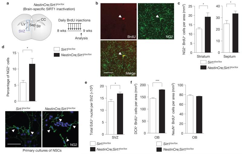
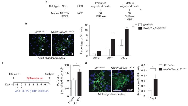
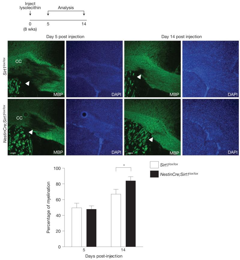
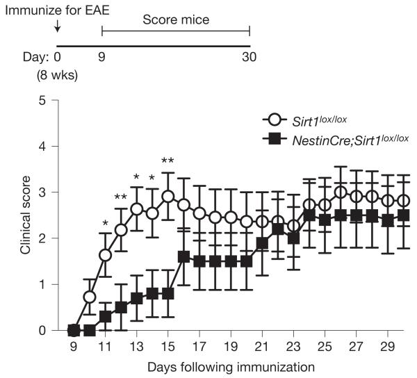
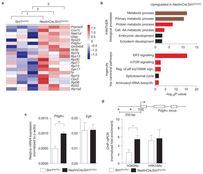
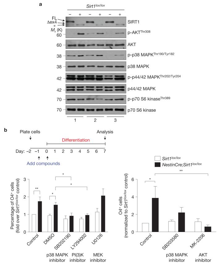
Oligodendrocytes-the myelin-forming cells of the central nervous system-can be regenerated during adulthood. In adults, new oligodendrocytes originate from oligodendrocyte progenitor cells (OPCs), but also from neural stem cells (NSCs). Although several factors supporting oligodendrocyte production have been characterized, the mechanisms underlying the generation of adult oligodendrocytes are largely unknown. Here we show that genetic inactivation of SIRT1, a protein deacetylase implicated in energy metabolism, increases the production of new OPCs in the adult mouse brain, in part by acting in NSCs. New OPCs produced following SIRT1 inactivation differentiate normally, generating fully myelinating oligodendrocytes. Remarkably, SIRT1 inactivation ameliorates remyelination and delays paralysis in mouse models of demyelinating injuries. SIRT1 inactivation leads to the upregulation of genes involved in cell metabolism and growth factor signalling, in particular PDGF receptor α (PDGFRα). Oligodendrocyte expansion following SIRT1 inactivation is mediated at least in part by AKT and p38 MAPK-signalling molecules downstream of PDGFRα. The identification of drug-targetable enzymes that regulate oligodendrocyte regeneration in adults could facilitate the development of therapies for demyelinating injuries and diseases, such as multiple sclerosis.


Figures

Figure 1
Figure 1
SIRT1 is expressed in adult NSCs and neural progenitors. (a) SIRT1 is expressed in the nuclei of NESTIN-positive cells in adult NSC and neural progenitor cultures. Immunocytochemistry on adherent monolayer cultures of adult NSCs and neural progenitors using antibodies against SIRT1 (green) and against the NSC/progenitor marker NESTIN (red). DAPI (blue) is a nuclear marker. (b) SIRT1 is expressed in NSCs and neural progenitors in the SVZ of the adult brain. Immunohistochemistry on brain sections of 8-week-old mice using antibodies against SIRT1 (green) and SOX2 (red, upper panel) or Ki67 (red, lower panel). White arrowheads show examples of double-stained cells. Insets show magnification of a cell positive for SIRT1 and SOX2 or Ki67. Merged images include DAPI to illustrate nuclei. Scale bars, 50 μm.
Figure 2
Figure 2
Inducible inactivation of SIRT1 in adult NSCs and neural progenitors expands the oligodendrocyte lineage. (a) SIRT1 can be conditionally inactivated in adult NSCs and neural progenitors by tamoxifen injection of NestinCreER;mT/mG;Sirt1lox/lox mice. Upper left: schematic of a coronal mouse brain section. LV, lateral ventricle; SVZ, subventricular zone; Sep, septum; Str, striatum; CC, corpus callosum. Not drawn to scale. Upper right, adult mice were injected with tamoxifen once before analysis. Bottom, membrane-targeted green fluorescent protein (mGFP) serves as a reporter for Cre recombinase activity in NestinCreER;mT/mG;Sirt1lox/lox mice. (b) Progeny of adult NSCs and neural progenitors were observed in the striatum and septum of both NestinCreER;mT/mG;Sirt1lox/lox and NestinCreER;mT/mG control mice 10 days after tamoxifen injection. Concentrated areas of membrane-targeted tomato red fluorescent protein (mTFP) are blood vessels. (c) Inactivation of SIRT1 in adult NSCs and neural progenitors leads to greater production of OLIG2-positive NSC and neural progenitor progeny in the surrounding NSC niche. Mean±s.e.m. of 4 independent cohorts of 2 mice per genotype for each cohort, except one in which there were 3 control mice (n = 4). Two-tailed, paired Student’s t-test, striatum, *P =0.0294; septum, *P =0.0174. Arrowheads point to cells co-expressing mGFP and OLIG2. (d) mGFP-positive cells with morphologies characteristic of myelinating oligodendrocytes were observed in the striatum and septum of NestinCreER;mT/mG;Sirt1lox/lox brains. (e) Deletion of active SIRT1 in adult NSCs and neural progenitors leads to greater production of GFAP-positive NSC and neural progenitor progeny in the surrounding NSC niche. Mean±s.e.m. of 4 independent cohorts of 2 mice per genotype for each cohort, except one in which there were 3 control mice (n = 4). Two-tailed, paired Student’s t-test, striatum, *P = 0.0282; septum, *P = 0.0144. Arrowheads point to cells co-expressing mGFP and GFAP. (f) Deletion of active SIRT1 in adult NSCs and neural progenitors enhances the area in the olfactory bulb (OB) covered by mGFP fluorescence 10 days after tamoxifen injection. Mean±s.e.m. of n = 3 NestinCreER;mT/mG and n = 4 NestinCreER;mT/mG;Sirt1lox/lox mice. Two-tailed, unpaired Student’s t-test, *P = 0.01. Tam, tamoxifen. All scale bars, 50 μm.
Figure 3
Figure 3
Brain-specific SIRT1 inactivation amplifies the proliferating OPC and NSC populations. (a) Mice lacking active SIRT1 specifically in NSCs and neural progenitors during embryonic development were generated by breeding mice in which exon 4 of Sirt1 is flanked by loxP sites (Sirt1lox/lox) with mice carrying a transgene in which Cre recombinase is expressed under the Nestin promoter (NestinCre). NestinCre;Sirt1lox/lox and control Sirt1lox / lox adult mice were injected with bromodeoxyuridine (BrdU) for 7 days to assess proliferation. SVZ, subventricular zone; LV, lateral ventricle; Sep, septum; Str, striatum; CC, corpus callosum. Schematic not drawn to scale. (b) Proliferating NG2-positive cells were observed in the striatum and septum of both NestinCre;Sirt1lox/lox and Sirt1lox/lox mice injected with BrdU for 7 days. Arrowheads indicate a double-labelled cell that is a dividing OPC. Scale bar, 50 μm. (c) The density of proliferating OPCs in the striatum and septum is greater in NestinCre;Sirt1lox/lox mice when compared with control mice. Mean±s.e.m. of n = 5 Sirt1lox/lox and n 4 = NestinCre;Sirt1lox/lox mice. Two-tailed, unpaired Student’s t-test; striatum, *P = 0.0139; septum, P = 0.041. (d) Absence of active SIRT1 increases the percentage of NG2-positive cells in proliferating NSC and neural progenitor cultures. Primary NSC and neural progenitor cultures from Sirt1lox/lox and NestinCre;Sirt1lox/lox 8-week-old mice were grown for a week in proliferation medium, and then immunostained for the OPC marker NG2. Arrowheads indicate NG2-positive cells. Mean±s.e.m. of 4 independent experiments. Two-tailed, unpaired Student’s t-test, *P = 0.0209. Scale bar, 50 μm. (e) Adult mice lacking SIRT1 in the nervous system have increased numbers of proliferating cells in the SVZ. Mice were injected with ethynyldeoxyuridine (EdU) for 2 h. Mean±s.e.m. of 4 mice per genotype. Two-tailed, unpaired Student’s t-test, *P = 0.0257. (f) The density of DCX-positive proliferating neuroblasts, but not NeuN-positive newly formed neurons, in the olfactory bulb (OB) granule cell layer is greater in NestinCre;Sirt1lox/lox mice when compared with control Sirt1lox/lox mice. Mice were injected with BrdU daily for 7 days before analysis. Mean±s.e.m. of 3 mice per genotype. Two-tailed, unpaired Student’s t-test, ***P = 0.0009.
Figure 4
Figure 4
SIRT1 inactivation does not alter the differentiation and myelination potential of oligodendrocytes. (a) Markers for specific cell types during differentiation of NSCs into mature oligodendrocytes. (b) Absence of active SIRT1 increases the percentage of O4-positive cells in differentiated adult NSC and neural progenitor cultures. Primary NSC and neural progenitor cultures from Sirt1lox/lox and NestinCre;Sirt1lox/lox 8-week-old mice were differentiated by growth factor removal and addition of 1% fetal bovine serum (FBS) after the first passage and immunostained after 2, 4 and 7 days for the oligodendrocyte marker O4. Images are representative of O4-positive cells at day 7 of differentiation. Arrowheads indicate individual O4-positive cells. Mean±s.e.m. of 5 independent experiments. Two-way ANOVA analysis: both genotype and differentiation day are significantly different, **P = 0.0055 and P = 0.0029, respectively. No interaction. Bonferroni post-test: day 7, P < 0.05. (c) SIRT1 inhibition enhances production of oligodendrocytes from NSCs and neural progenitor cells. Proliferating NSCs and neural progenitors were incubated with the SIRT1-specific inhibitor EX-527 (5 μM) or dimethylsulphoxide (DMSO) vehicle for 24 h. Cells were then switched to differentiation media (1% FBS) containing the same concentration of compound or vehicle and grown for a further 48 h. Cells were allowed to differentiate without added compound for 5 more days before staining for the oligodendrocyte marker O4. Mean±s.e.m. of 3 independent experiments. One-tailed, unpaired, Student’s t-test, *P < 0.05. (d) Absence of active SIRT1 increases the percentage of MBP-positive cells in differentiated adult NSC and neural progenitor cultures. Primary NSC and neural progenitor cultures from Sirt1lox/lox and NestinCre;Sirt1lox/lox 8-week-old mice were differentiated for 7 days by growth factor removal and addition of 1% FBS after the first passage and immunostained 7 days for the mature oligodendrocyte marker MBP. Images are representative of MBP-positive cells at day 7 of differentiation. Mean±s.e.m. of 6 independent experiments. Two-tailed, unpaired Student’s t-test, *P = 0.0398. Scale bars, 50 μm (b,d).
Figure 5
Figure 5
SIRT1 inactivation enhances remyelination in the corpus callosum after lysolecithin-induced demyelination. Mice lacking active SIRT1 in the nervous system exhibit enhanced remyelination after focal lysolecithin-induced demyelination in the corpus callosum (CC). Sirt1lox/lox and NestinCre;Sirt1lox/lox 8-week-old mice were stereotactically injected with 1% lysolecithin in the corpus callosum and allowed to recover for 5 or 14 days. Levels of myelination were blindly quantified in the region of injection by coverage of MBP staining over background. One-tailed, unpaired Student’s t-test, *P = 0.028. Mean±s.e.m. of n = 3 Sirt1lox/lox and n = 5 NestinCre;Sirt1lox/lox mice for day 5 and n = 10 Sirt1lox/lox and n = 8 NestinCre;Sirt1lox/lox mice for day 14. Arrowheads indicate demyelination areas. Scale bars, 50 μm.
Figure 6
Figure 6
SIRT1 inactivation delays onset of paralysis in chronic EAE. Mice lacking active SIRT1 in the nervous system are initially protected against the onset of paralysis in EAE. Mice were assessed for clinical signs of EAE daily after immunization with MOG35–55 peptide, Freund’s adjuvant and pertussis toxin. Score of 0, no paralysis; 1, loss of tail tone; 2, hindlimb weakness; 3, hindlimb paralysis; 4, hindlimb and forelimb paralysis; 5, moribund or dead. Mean±s.e.m. of n = 11 Sirt1lox/lox and n = 10 NestinCre;Sirt1lox/lox mice. Mann–Whitney U test, *P < 0.05, **P < 0.01.
Figure 7
Figure 7
SIRT1 inactivation leads to the upregulation of genes involved in metabolism, protein translation and growth factor signalling. (a) Genes upregulated in NestinCre;Sirt1lox/lox NSCs and neural progenitors compared with Sirt1lox/lox control NSCs and neural progenitors. Whole-genome microarray data from three independent biological replicates of proliferating NestinCre;Sirt1lox/lox and Sirt1lox/lox NSCs and neural progenitor isolated from 8-week-old mice. Heat map represents a subset of genes upregulated in the absence of active SIRT1 that was selected on the basis of a false discovery rate of less than 0.001%. Numbers 1–3 designate independent biological experiments. Colours represent the Z score of the robust multi-array analysis-normalized expression level for each gene (red is high expression; blue is low expression). Asterisk highlights Pdgfrα, a gene important for OPC proliferation and maintenance,,,. (b) Genes upregulated following SIRT1 inactivation are significantly over-represented in biological processes related to metabolism and ribosomal biology. Genes differentially regulated by SIRT1 were selected on the basis of a false discovery rate of less than 1% and analysed using PANTHER and Ingenuity Pathway Analysis (IPA). Canonical pathways significantly over-represented in the genes upregulated following SIRT1 inactivation are presented. Upper graph shows 6 of the top 7 PANTHER biological processes. Lower graph shows the top 5 IPA processes. Dark red colour highlights categories related to metabolism and bright red colour highlights categories related to protein synthesis and metabolism. AA, amino acid. (c) Pdgfrα mRNA expression is greater in proliferating NSCs and neural progenitors lacking active SIRT1 than in control cells. RT–qPCR on total RNA from primary NSC and neural progenitor cultures from NestinCre;Sirt1lox / lox and Sirt1lox/lox 8-week-old mice with primers to Pdgfrα and Egfr. Expression of Pdgfrα and Egfr was normalized to β-actin. Mean±s.e.m. of 3 independent experiments. Two-tailed, unpaired Student’s t-test, **P = 0.0068. (d) Increased H3K9 acetylation at the Pdgfrα promoter in NSCs and neural progenitors lacking active SIRT1. ChIP-qPCR on NSCs and neural progenitors from Sirt1lox / lox and NestinCre;Sirt1lox/lox 8-week-old mice with antibodies against H3K9Ac or H4K16Ac with primers to the Pdgfrα promoter. Data are presented as fold enrichment at the Pdgfrα promoter normalized to that at the promoter of HoxA10, a silent gene in these cells. Mean±s.e.m. of 3 independent experiments. Two-tailed, unpaired Student’s t-test, *P = 0.0491. TSS, transcriptional start site. The Pdgfrα locus is not drawn to scale.
Figure 8
Figure 8
Inhibition of p38 MAPK and AKT signalling reduces the production of oligodendrocytes from NSCs and neural progenitors lacking active SIRT1. (a) Increased AKT and p38 MAPK phosphorylation in NSCs and neural progenitors lacking active SIRT1. Immunoblots of lysates from 3 independent cultures of NSC and neural progenitors isolated from Sirt1lox / lox and NestinCre;Sirt1lox / lox adult mice. Antibodies specific against phosphorylated forms of AKT, p38 MAPK, p44/42 MAPK and p70 S6 kinase were used. Antibodies specific against total proteins were used as loading controls. FL, full-length SIRT1; Δex4, truncated inactive SIRT1 protein generated from the Sirt1 gene that lacks exon 4. *, protein that is distinct from the truncated SIRT1 protein and probably represents a splice variant of SIRT1 (see Supplementary Fig. S2d). (b) Activity of signalling kinases p38 MAPK and AKT is important for oligodendrocyte generation from NSCs and neural progenitors. Proliferating Sirt1lox / lox and NestinCre;Sirt1lox/lox NSCs and neural progenitors were incubated with p38 MAPK inhibitor SB202190 (10 μM) or SB203580 (1 μM), PI(3)K inhibitor LY294002 (10 μM), AKT inhibitor MK-2206 (1 μm), MEK inhibitor U0126 (10 μm) or dimethylsulphoxide (DMSO) vehicle for 24 h. Cells were then switched to differentiation medium (1% FBS) containing the same concentration of compounds or vehicle and grown for a further 48 h. Cells were allowed to differentiate without added compounds for 5 more days, with complete medium changes every other day. Cells were stained for the oligodendrocyte marker O4. Left panel: mean±s.e.m. of the following number of independent experiments, n = 9 for control and dimethylsulphoxide; n = 5 for SB202190; n = 6 for LY294002; and n = 4 for U0126. Two-way ANOVA, compound, P < 0.0001; genotype, P < 0.0001; interaction, P = 0.0165; Bonferroni post-tests, *P < 005, **P < 0.01. Right panel: mean ± s.e.m. of 4 independent experiments. Two-way ANOVA, compound, P = 0.0684; genotype, P = 0.0438, interaction, P = 0.0416; Bonferroni post-tests, *P < 0.05, **P < 0.01. = Uncropped images of blots are shown in Supplementary Fig. S8.

Comment in

Similar articles

Cited by

References

    1. McTigue DM, Tripathi RB. The life, death, and replacement of oligodendrocytes in the adult CNS. J. Neurochem. 2008;107:1–19. - PubMed
    1. Franklin RJ, Ffrench-Constant C. Remyelination in the CNS: from biology to therapy. Nat. Rev. Neurosci. 2008;9:839–855. - PubMed
    1. Picard-Riera N, et al. Experimental autoimmune encephalomyelitis mobilizes neural progenitors from the subventricular zone to undergo oligodendrogenesis in adult mice. Proc. Natl Acad. Sci. USA. 2002;99:13211–13216. - PMC - PubMed
    1. Nait-Oumesmar B, et al. Progenitor cells of the adult mouse subventricular zone proliferate, migrate and differentiate into oligodendrocytes after demyelination. Eur. J. Neurosci. 1999;11:4357–4366. - PubMed
    1. Menn B, et al. Origin of oligodendrocytes in the subventricular zone of the adult brain. J. Neurosci. 2006;26:7907–7918. - PMC - PubMed

Publication types

MeSH terms

Substances