. 2013 May;8(5):477-85.
doi: 10.4161/epi.24547. Epub 2013 Apr 17.

Large-scale global identification of protein lysine methylation in vivo

Affiliations

Large-scale global identification of protein lysine methylation in vivo

Xing-Jun Cao et al. Epigenetics. 2013 May.

Abstract

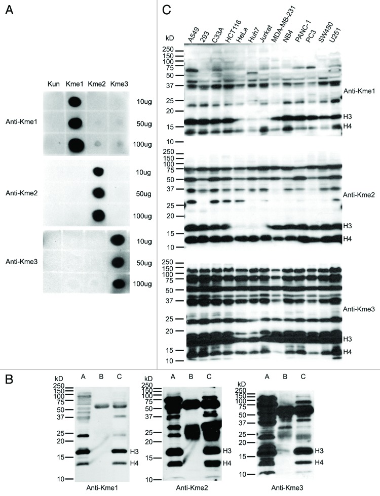
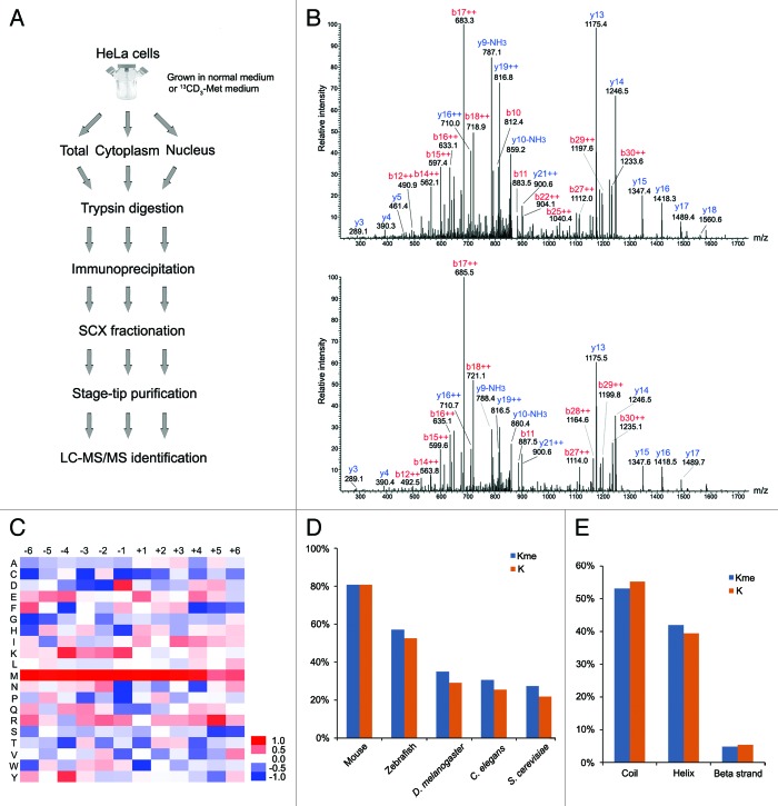
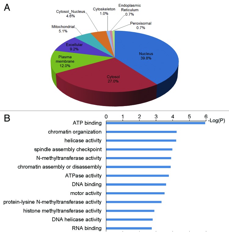
Lysine methylation mediated by methyltransferase enzymes is present on multiple proteins throughout the cell; however, methods to uncover and characterize global protein lysine methylation patterns do not readily exist. Here we developed pan-specific methyl lysine antibodies that we utilized in immunoprecipitation experiments coupled with mass spectrometry to yield one of the first large-scale surveys of protein lysine methylation in vivo. In total, 552 different lysine methylation sites were determined, making this one of the most comprehensive global studies published to date. The large majority of these sites have not been yet reported. These sites showed significantly enriched sequence motifs and resided in proteins that are involved in diverse biological processes, particularly in chromatin organization. Our data provide a comprehensive view of lysine methylation in human cells and a powerful resource to facilitate investigations into the function of lysine methylation on non-histone proteins.

Keywords: affinity; histone; immunoprecipitation; mass spectrometry; methylation; non-histone; proteomics.


Figures

None
Figure 1. Characterization of the antibodies against mono-, di- and tri-methyl lysine. (A) Dot blot analysis of the pan anti-methyl lysine antibodies. Peptides with un- (Kun), mono- (Kme1), di- (Kme2) and tri- (Kme3) methyl lysine were blotted onto nitrocellulose membranes and probed with the antibodies, respectively. (B) Immunoprecipitation (IP) analysis of lysates prepared from HeLa cells using the antibodies. Lanes A, B and C are input, IP eluates with normal rabbit IgG (negative control) and IP eluates with anti-methyl lysine antibodies, respectively. The concentration of SDS-PAGE was 15%. (C) Lysine methylation profiles in 13 human cell lines. The protein lysates of 13 cell lines were separated by 12.5% SDS-PAGE, transferred to PVDF membranes and probed with the antibodies, respectively.
None
Figure 2. Illustration of the study strategy and characterization of the methylation sites. (A) Illustration of the study strategy. HeLa cells cultured in normal or 13CD3-methionine medium were lysed, or sub-cellularly fractionated to separate cytoplasm and nucleus, and their protein lysates were digested by trypsin. The methylated peptides were enriched through immunoprecipitation and underwent strong cation-exchange (SCX) separation. Each fraction was desalted and subjected to mass spectrometry. (B) MS/MS spectra of the methylated peptide, TKme1AAAAAAAAAPAAAATAPTTAATTAATAAQ. The upper and lower spectra come from normal and heavy methyl-labeled samples, respectively. (C) Heat map of relative distribution of amino acids around the methylated lysine sites. Amino acids from -6 to +6 positions adjacent to the methylated lysine sites and all lysine residues were extracted, respectively. At each position, the frequency of amino acid around methylated lysine sites was divided by that around all lysine residues, and the ratios were visually represented after logarithmic transformation (log2). Color scale reflects the degree of enrichment (red) or depletion (blue). (D) Evolutionary conservation of the methylated lysine sites and all lysines throughout the eukaryotic species. “Kme” and “K” denote methylated lysines and all lysines, respectively. (E) Distribution of methylated lysines and all lysines in protein secondary structure. “Kme” and “K” denote methylated lysines and all lysines, respectively.
None
Figure 3.(A) Subcellular localization of methylated proteins. (B) Over-represented GO terms of methylated proteins.

Similar articles

Cited by

References

    1. Kouzarides T. Histone methylation in transcriptional control. Curr Opin Genet Dev. 2002;12:198–209. doi: 10.1016/S0959-437X(02)00287-3. - DOI - PubMed
    1. Lachner M, Jenuwein T. The many faces of histone lysine methylation. Curr Opin Cell Biol. 2002;14:286–98. doi: 10.1016/S0955-0674(02)00335-6. - DOI - PubMed
    1. Sims RJ, 3rd, Nishioka K, Reinberg D. Histone lysine methylation: a signature for chromatin function. Trends Genet. 2003;19:629–39. doi: 10.1016/j.tig.2003.09.007. - DOI - PubMed
    1. Greer EL, Shi Y. Histone methylation: a dynamic mark in health, disease and inheritance. Nat Rev Genet. 2012;13:343–57. doi: 10.1038/nrg3173. - DOI - PMC - PubMed
    1. Simon JA, Lange CA. Roles of the EZH2 histone methyltransferase in cancer epigenetics. Mutat Res. 2008;647:21–9. doi: 10.1016/j.mrfmmm.2008.07.010. - DOI - PubMed

Publication types

LinkOut - more resources