. 2013 May 15;73(10):3145-54.
doi: 10.1158/0008-5472.CAN-13-0011. Epub 2013 May 3.

β1 integrin targeting potentiates antiangiogenic therapy and inhibits the growth of bevacizumab-resistant glioblastoma

Affiliations

β1 integrin targeting potentiates antiangiogenic therapy and inhibits the growth of bevacizumab-resistant glioblastoma

W Shawn Carbonell et al. Cancer Res. .

Abstract

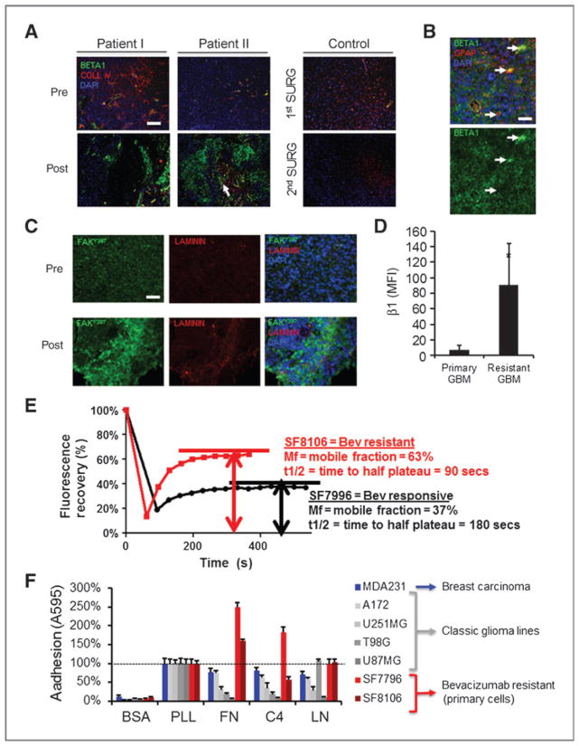
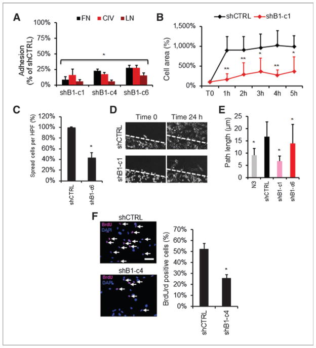
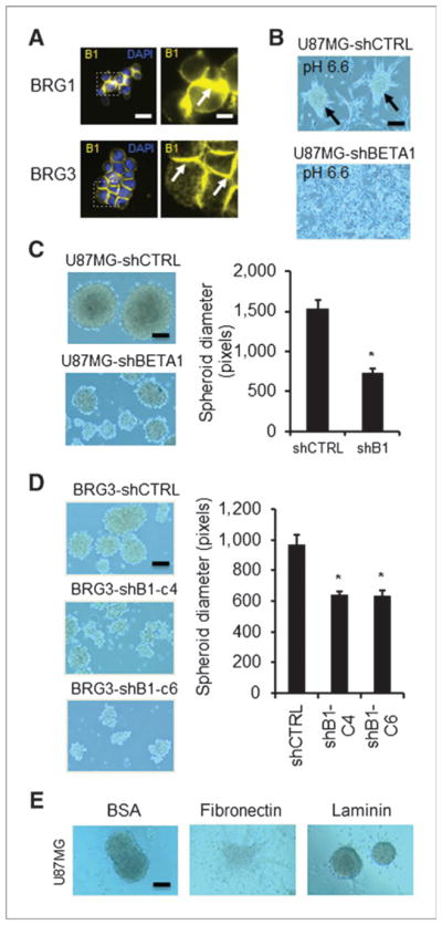
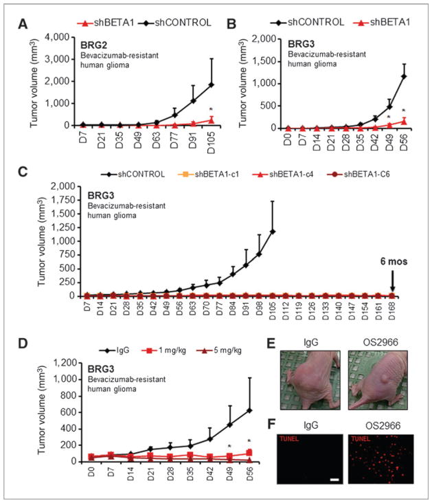
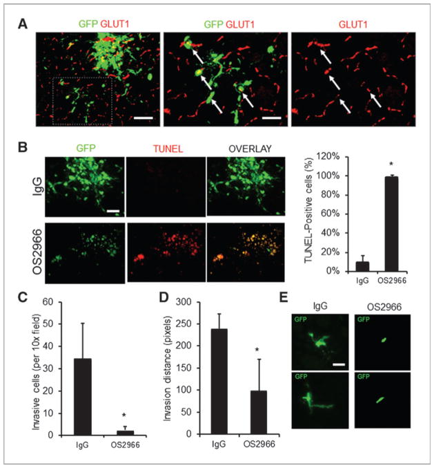
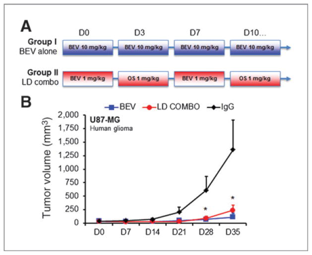
Antiangiogenic therapies like bevacizumab offer promise for cancer treatment, but acquired resistance, which often includes an aggressive mesenchymal phenotype, can limit the use of these agents. Upregulation of β1 integrin (ITGB1) occurs in some bevacizumab-resistant glioblastomas (BRG) whereby, mediating tumor-microenvironment interactions, we hypothesized that it may mediate a mesenchymal-type resistance to antiangiogenic therapy. Immunostaining analyses of β1 integrin and its downstream effector kinase FAK revealed upregulation in 75% and 86% of BRGs, respectively, compared with pretreatment paired specimens. Furthermore, flow cytometry revealed eight-fold more β1 integrin in primary BRG cells compared with cells from bevacizumab-naïve glioblastomas (BNG). Fluorescence recovery after photobleaching of cells engineered to express a β1-GFP fusion protein indicated that the mobile β1 integrin fraction was doubled, and half-life of β1 integrin turnover in focal adhesions was reduced markedly in BRG cells compared with bevacizumab-responsive glioblastoma multiforme cells. Hypoxia, which was increased with acquisition of bevacizumab resistance, was associated with increased β1 integrin expression in cultured BNG cells. BRGs displayed an aggressive mesenchymal-like phenotype in vitro. We found that growth of BRG xenograft tumors was attenuated by the β1 antibody, OS2966, allowing a 20-fold dose reduction of bevacizumab per cycle in this model. Intracranial delivery of OS2966 through osmotic pumps over 28 days increased tumor cell apoptosis, decreased tumor cell invasiveness, and blunted the mesenchymal morphology of tumor cells. We concluded that β1 integrin upregulation in BRGs likely reflects an onset of hypoxia caused by antiangiogenic therapy, and that β1 inhibition is well tolerated in vivo as a tractable strategy to disrupt resistance to this therapy.


Conflict of interest statement

Disclosure of Potential Conflicts of Interest

W. Shawn Carbonell is employed as a cofounder, President, and CEO, and has ownership interest (including patents) in OncoSynergy, Inc. C.C. Park has ownership interest (including patents) in Oncosynergy Ltd. M.K. Aghi is employed as a scientific advisory board member/shareholder, has ownership interest (including patents), and is a consultant/advisory board member of Oncosynergy. No potential conflicts of interest were disclosed by the other authors.

Figures

Figure 1
Figure 1
β1 integrin expression and bevacizumab resistance in glioblastoma. A, immunofluorescent staining for β1 integrin (green); collagen IV, an endothelial marker (red); and 4, 6-diamidino-2-phenylindole nuclear counterstain in clinical specimens from 2 patients before and after development of bevacizumab resistance and a control patient at first and second surgery. Scale bar, 100 μm. B, high-powered view from another BRG shows β1 integrin immunoreactivity in glioma cells including glial fibrillary acidic protein-positive cells (arrows). Scale bar, 50 μm. C, phosphorylated focal adhesion kinase (FAKY397, green) is enriched in post-bevacizumab tissues and correlated with ECM consistent with increased integrin activity. Scale bar, 120 μm. D, dissociated cells from resistant glioblastoma (GBM) have significantly higher β1 integrin on flow cytometry than primary tumor cells (*, P < 0.05, t test). E, FRAP conducted on cells expressing β1-GFP fusion protein revealed more rapid β1 integrin turnover in focal adhesions and a higher integrin mobile fraction in bevacizumab-resistant SF8106 cells than in bevacizumab-responsive SF7996 cells. Experiment was repeated 4 times, with distinct curves each time (P < 0.001, t test). F, BRG cells are more adherent to classical ECM proteins than several glioblastoma cell lines in addition to the highly adherent MDA-MB-231 breast carcinoma (BSA, negative control; PLL, positive control; FN, fibronectin; C4, collagen type IV; LN, laminin). Data are mean ± SD. See also Supplementary Fig. S2.
Figure 2
Figure 2
β1 integrin knockdown in resistant glioblastoma cells inhibits mesenchymal function. β1 integrin knockdown clones are impaired in cell adhesion (A; *, P < 0.01, ANOVA with post hoc Dunnett test), dynamic cell spreading (B; *, P < 0.05, **, P < 0.01, ANOVA with post hoc Dunnett test), and percentage of spread cells (C; *, P < 0.01, Mann–Whitney test) on fibronectin. D and E, dynamic time-lapse analysis in a scratch wound assay shows impaired migration in knockdown clones versus control (*, P < 0.05, ANOVA with post hoc Dunnett test). β1 integrin knockdowns show decreased proliferation as shown by BrdU immunofluorescence (F; *, P < 0.01, t test). Scale bar, 120 μm. Data are mean ± SD. See also Supplementary Figs. S3 to S5.
Figure 3
Figure 3
β1 knockdown in glioma cells inhibits spheroidal growth. A, immunofluorescence shows acutely enriched β1 immunoreactivity at cell–cell contacts in 2 BRG lines in an in vitro aggregation assay. Scale bars, 30 and 10 μm, respectively. B, β1 knockdown in U87MG attenuates spheroid formation (arrows) in acidic media. Scale bar, 120 μm. Formal spheroidal growth assay confirms impairment of spheroid diameter with β1 knockdown in both U87MG (C; *, P < 0.05, t test; scale bar, 120 μm) and BRG3 clones (D; *, P < 0.05, ANOVA with post-hoc Dunnett test; scale bar, 120 μm). E, U87MG spheroids unravel when plated on fibronectin, but not BSA control substrate consistent with a β1-mediated mechanism. Scale bar, 120 μm. Data are mean ± SD. See also Supplementary Fig. S6.
Figure 4
Figure 4
β1 integrin inhibition in BRG cells attenuates growth in vivo. Growth of polyclonal β1 integrin knockdowns in a subcutaneous xenograft model was attenuated in both BRG2 (A; *, P < 0.05, ANOVA with post hoc Bonferroni test) and BRG3 (B; *, P < 0.05, ANOVA with post hoc Bonferroni test) lines. C, more than 90% knockdown of β1 integrin in BRG3 clones prevents in vivo growth for up to 6 months. D and E, inhibitory anti-integrin antibody, OS2966, similarly inhibits growth of BRG3 cells in vivo at 1 or 5 mg/kg i.p. biweekly compared with control IgG (*, P < 0.05, ANOVA with post hoc Bonferroni test). F, 5 mg/kg of OS2966 induces more apoptosis in subcutaneous tumors than IgG. Scale bar, 100 μm. Data are mean ± SEM. See also Supplementary Fig. S7A.
Figure 5
Figure 5
OS2966 inhibits growth and invasion of orthotopically implanted BRG3 cells. A, low-power epifluorescent microscopy shows parenchymal invasion of BRG3 cells in IgG-treated control mice. Many instances of this invasion (>50%) occurred perivascularly consistent with vessel cooption (arrows). Scale bar, 120 μm. B, induction of apoptosis after OS2966 versus IgG control mAb treatment (*, P < 0.05, t test). Scale bar, 120 μm. Invasion of BRG3 cells in OS2966-treated mice was significantly attenuated as shown by number of invasive cells (C; *, P < 0.05, t test), invasion distance (D; *, P < 0.05, t test), and was supported by altered morphology of single invading cells including length and number of cellular processes (E). Scale bar, 50 μm. Data are mean ± SD. See also Supplementary Fig. S7.
Figure 6
Figure 6
OS2966 potentiates efficacy of bevacizumab in a glioma model. A, study design of experimental groups with standard bevacizumab therapy at 10 mg/kg biweekly (group I, BEV) versus alternating bevacizumab and OS2966 at 1 mg/kg (group II, low-dose combo). B, inhibition of tumor growth with low-dose combo (group II) was equivalent to high-dose bevacizumab (group I) compared with IgG controls in subcutaneous U87MG xenografts (*, P < 0.05, ANOVA with post hoc Bonferroni test). Shown are mean ± SEM.

Similar articles

Cited by

References

    1. Stupp R, Dietrich PY, Ostermann Kraljevic S, Pica A, Maillard I, Maeder P, et al. Promising survival for patients with newly diagnosed glioblastoma multiforme treated with concomitant radiation plus temozolomide followed by adjuvant temozolomide. J Clin Oncol. 2002;20:1375–82. - PubMed
    1. Kreisl TN, Kim L, Moore K, Duic P, Royce C, Stroud I, et al. Phase II trial of single-agent bevacizumab followed by bevacizumab plus irinotecan at tumor progression in recurrent glioblastoma. J Clin Oncol. 2009;27:740–5. - PMC - PubMed
    1. Kienast Y, Winkler F. Therapy and prophylaxis of brain metastases. Expert Rev Anticancer Ther. 2010;10:1763–77. - PubMed
    1. Vredenburgh JJ, Desjardins A, Herndon JE, II, Dowell JM, Reardon DA, Quinn JA, et al. Phase II trial of bevacizumab and irinotecan in recurrent malignant glioma. Clin Cancer Res. 2007;13:1253–9. - PubMed
    1. Gotzmann J, Mikula M, Eger A, Schulte-Hermann R, Foisner R, Beug H, et al. Molecular aspects of epithelial cell plasticity: implications for local tumor invasion and metastasis. Mutat Res. 2004;566:9–20. - PubMed

Publication types