. 2013 Jun;20(6):687-95.
doi: 10.1038/nsmb.2562. Epub 2013 May 5.

The octamer is the major form of CENP-A nucleosomes at human centromeres

Affiliations

The octamer is the major form of CENP-A nucleosomes at human centromeres

Dan Hasson et al. Nat Struct Mol Biol. 2013 Jun.

Abstract

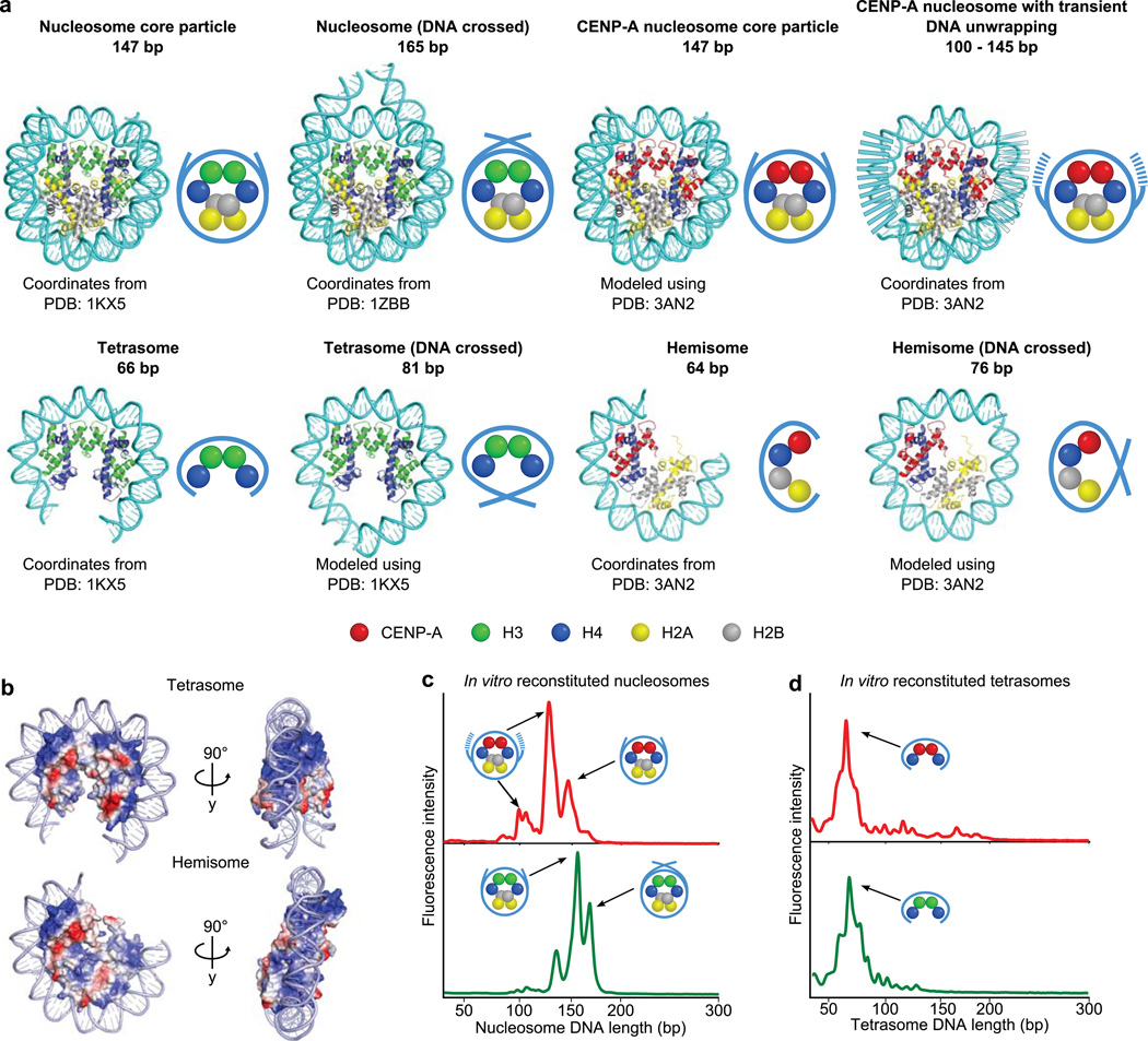
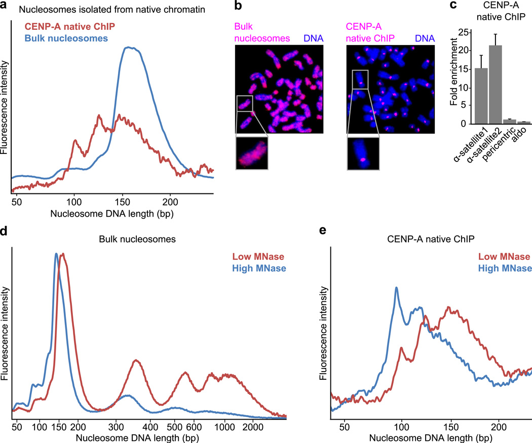
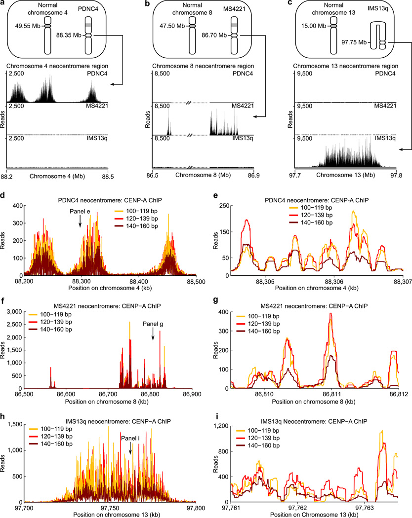
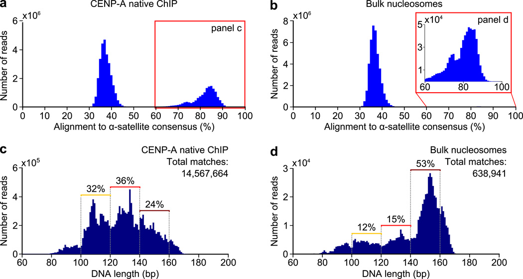
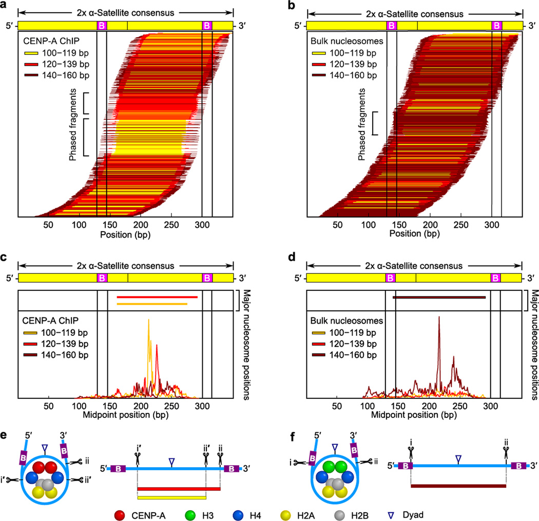
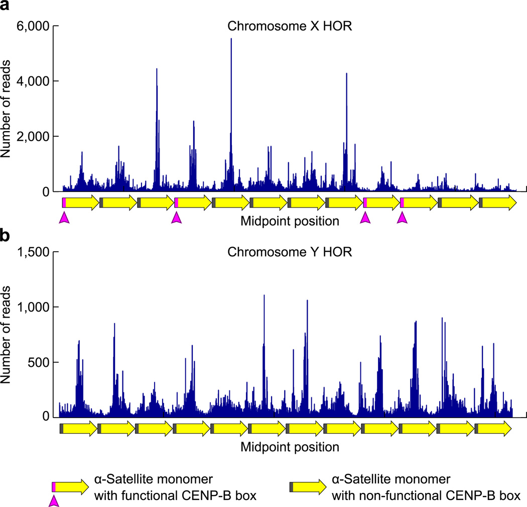
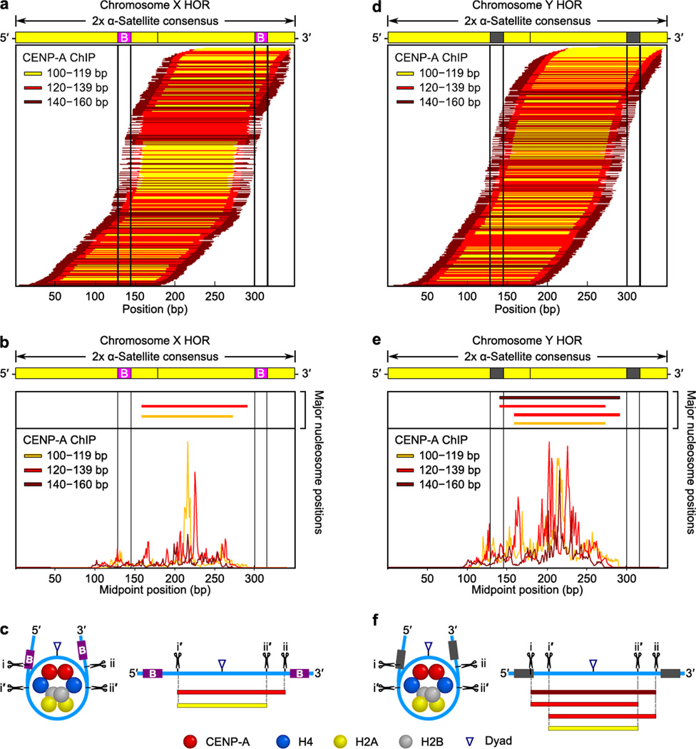
The centromere is the chromosomal locus that ensures fidelity in genome transmission at cell division. Centromere protein A (CENP-A) is a histone H3 variant that specifies centromere location independently of DNA sequence. Conflicting evidence has emerged regarding the histone composition and stoichiometry of CENP-A nucleosomes. Here we show that the predominant form of the CENP-A particle at human centromeres is an octameric nucleosome. CENP-A nucleosomes are very highly phased on α-satellite 171-base-pair monomers at normal centromeres and also display strong positioning at neocentromeres. At either type of functional centromere, CENP-A nucleosomes exhibit similar DNA-wrapping behavior, as do octameric CENP-A nucleosomes reconstituted with recombinant components, having looser DNA termini than those on conventional nucleosomes containing canonical histone H3. Thus, the fundamental unit of the chromatin that epigenetically specifies centromere location in mammals is an octameric nucleosome with loose termini.


Figures

Figure 1
Figure 1. Structure-based predictions for MNase protection and experimental outcomes with CENP-A-containing particles assembled with recombinant components
(a) Molecular models (cartoon representations, right) of the indicated, proposed DNA–protein particles demonstrating the expected length of DNA protected following MNase digestion. (b) Electrostatic surface potential maps depicting the predicted path of DNA wrapping a CENP-A-containing tetrasome or hemisome, where positively charged surfaces are colored in blue and negatively charged surfaces are colored in red. Note that 64–66 bp completely covers the DNA wrapping surface of either tetrameric configuration. (c,d) MNase digestion profiles of octameric CENP-A- or H3-containing mononucleosomes (c) or tetrasomes (d) reconstituted on a 200 bp template.
Figure 2
Figure 2. Nuclease digestion of native CENP-A-containing particles resembles that of octameric nucleosomes with loose termini
(a) DNA length distributions of MNase-digested CENP-A native ChIP and bulk nucleosomes from the same preparation. (b) Fluorescence in situ hybridization using DNA from bulk nucleosomes or CENP-A native ChIP as probes. Bulk nucleosome DNA labels the entire chromosome whereas CENP-A probe labels solely centromeric regions, as expected. (c) Quantitative real-time PCR analysis comparing enrichment of CENP-A native ChIP DNA relative to bulk nucleosome DNA. CENP-A ChIP sequences are enriched for α-satellite regions (α-satellite1, α-satellite2), but not at pericentric or promoter (aldo) regions, as expected. Error bars represent s.e.m. from three independent replicates. (d) Standard digestion (red) or overdigestion (blue, threefold higher concentration of MNase used) of chromatin. (e) DNA length distributions of CENP-A native ChIP following standard digestion (red) or overdigestion (blue) of chromatin.
Figure 3
Figure 3. The three size classes of CENP-A nucleosomes localize to the same prominent positions on neocentromeres
(a–c) Bowtie-mapped paired-end CENP-A native ChIP-Seq reads in three different human neocentromere-containing cell lines (PDNC4, MS4221, IMS13q; ideograms, top) demonstrate the specificity of CENP-A native ChIP. The IMS13q neocentromere was formed on an aberrant chromosome with an inversion duplication. The MS4221 neocentromere contains repetitive DNA denoted by dashed lines with strikethrough. (d–i) Occupancy maps for the three different size classes of CENP-A nucleosomes along the length of the neocentromere for PDNC4 (d), MS4221 (f), and IMS13q (h) and within a subsection (2500 bp window) in (e, g, and i).
Figure 4
Figure 4. CENP-A nucleosomes on the repetitive α-satellite DNA of normal centromeres have a tripartite distribution of nuclease protected DNA fragments
(a,b) Alignment of CENP-A (a) or bulk nucleosome (b) fragments to a dimer α-satellite consensus sequence. (c,d) Distribution of DNA lengths of all CENP-A (c) or bulk nucleosome (d) fragments aligning to the α-satellite consensus sequence with ≥ 60% identity.
Figure 5
Figure 5. Terminally unwrapped CENP-A nucleosomes and their conventional counterparts with wrapped termini are similarly phased at normal centromeres
(a,b) The position of each individual CENP-A (a) or bulk nucleosome (b) along a dimerized α-satellite consensus sequence is indicated by a horizontal line. Each fragment is color-coded based on length, as indicated. (c,d) The midpoint positions of CENP-A (c) or bulk nucleosome (d) fragments along the dimer α-satellite consensus sequence. Solid vertical lines indicate the location of the 17 bp CENP-B box (B) in (a–d). (e,f) Models of the preferred positioning and MNase cleavage sites on CENP-A (e) and bulk (f) nucleosomes at normal centromeres.
Figure 6
Figure 6. Phasing of CENP-A nucleosomes at annotated regions of α-satellite DNA from the X and Y chromosomes
(a,b) CENP-A nucleosome midpoint positions are shown along an annotated region of the 2 kb, 12 monomer HOR of α-satellite from the X chromosome (a) or a 12 monomer portion of the Y chromosome HOR (b).
Figure 7
Figure 7. CENP-A nucleosomes are less phased and gain symmetric MNase digestion on the Y chromosome centromere that lacks functional CENP-B boxes
(a–c) Maps of chromosome X HOR-aligned CENP-A sequences. (d–f). Maps of chromosome Y HOR-aligned CENP-A sequences. Data and models are shown in the same manner as for the global analysis of CENP-A nucleosome-associated α-satellite sequences (Fig. 5).

Comment in

Similar articles

Cited by

References

    1. Cleveland DW, Mao Y, Sullivan KF. Centromeres and kinetochores: from epigenetics to mitotic checkpoint signaling. Cell. 2003;112:407–421. - PubMed
    1. Mendiburo MJ, Padeken J, Fülöp S, Schepers A, Heun P. Drosophila CENH3 is sufficient for centromere formation. Science. 2011;334:686–690. - PubMed
    1. Barnhart MC, et al. HJURP is a CENP-A chromatin assembly factor sufficient to form a functional de novo kinetochore. J. Cell Biol. 2011;194:229–243. - PMC - PubMed
    1. Guse A, Carroll CW, Moree B, Fuller CJ, Straight AF. In vitro centromere and kinetochore assembly on defined chromatin templates. Nature. 2011;477:354–358. - PMC - PubMed
    1. Warburton PE. Chromosomal dynamics of human neocentromere formation. Chromosome Res. 2004;12:617–626. - PubMed

Methods References

    1. Carruthers LM, Tse C, Walker KP, Hansen JC. Assembly of defined nucleosomal and chromatin arrays from pure components. Meth. Enzymol. 1999;304:19–35. - PubMed
    1. Luger K, Rechsteiner TJ, Richmond TJ. Preparation of nucleosome core particle from recombinant histones. Meth. Enzymol. 1999;304:3–19. - PubMed
    1. Lowary PT, Widom J. New DNA sequence rules for high affinity binding to histone octamer and sequence-directed nucleosome positioning. J. Mol. Biol. 1998;276:19–42. - PubMed
    1. Huynh VAT, Robinson PJJ, Rhodes D. A Method for the In Vitro Reconstitution of a Defined ‘30nm’ Chromatin Fibre Containing Stoichiometric Amounts of the Linker Histone. J. Mol. Biol. 2005;345:957–968. - PubMed
    1. Brünger AT, et al. Crystallography & NMR system: A new software suite for macromolecular structure determination. Acta Crystallogr. D Biol. Crystallogr. 1998;54:905–921. - PubMed

Publication types

Associated data