

Oxygen tomography by Čerenkov-excited phosphorescence during external beam irradiation
- PMID: 23644902
- PMCID: PMC3643897
- DOI: 10.1117/1.JBO.18.5.050503
Oxygen tomography by Čerenkov-excited phosphorescence during external beam irradiation
Abstract
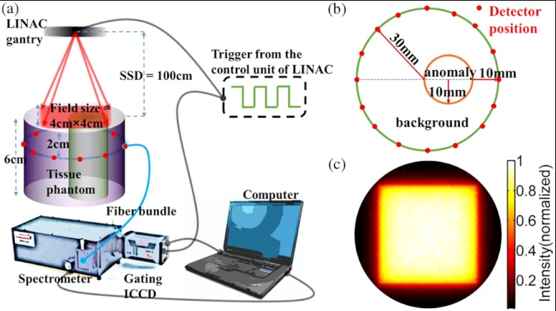
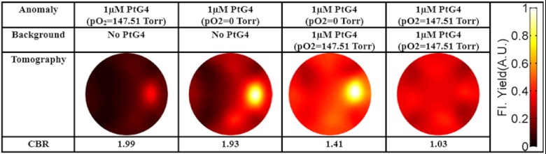
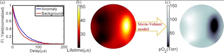
The efficacy of radiation therapy depends strongly on tumor oxygenation during irradiation. However, current techniques to measure this parameter in vivo do not facilitate routine monitoring in patients. Herein, we demonstrate a noninvasive method for tomographic imaging of oxygen partial pressure (pO(2)) in deep tissue using the phosphorescence decay of an oxygen-sensitive probe excited by Čerenkov radiation induced by external beam radiotherapy. Tissue-simulating scattering phantoms (60 mm diameter with a 20 mm anomaly) containing platinum(II)-G4 (PtG4), a dendritic porphyrin-based phosphor, whose phosphorescence is quenched in the presence of oxygen, were irradiated with a clinical linear accelerator. The emitted phosphorescence was measured at various positions on the phantom boundary using a spectrograph coupled to an intensified charge-coupled device (ICCD). At each position, PtG4 phosphorescence decay curves were measured by synchronizing the ICCD to the linear accelerator pulses. Tomographic images of phosphorescence yield and lifetime were recovered for phantoms with homogenous PtG4 concentrations and heterogeneous pO(2). Since PtG4 lifetime is strongly and predictably dependent on pO(2) through the Stern-Volmer relationship, tomographic images of pO(2) were also reported, and showed excellent agreement with independent oxygenation measurements. Translating this approach to the clinic could facilitate direct sensing of pO(2) during radiotherapy.
Figures
References
-
- Wilson D. F., Cerniglia G. J., “Localization of tumors and evaluation of their state of oxygenation by phosphorescence imaging,” Cancer Res. 52(14), 3988–3993 (1992). - PubMed
Publication types
MeSH terms
Substances
Grants and funding
LinkOut - more resources
Full Text Sources
Other Literature Sources
